. 2010 Dec 29;5(12):e15680.
doi: 10.1371/journal.pone.0015680.

Role of the chemokine receptors CCR1, CCR2 and CCR4 in the pathogenesis of experimental dengue infection in mice

Affiliations

Role of the chemokine receptors CCR1, CCR2 and CCR4 in the pathogenesis of experimental dengue infection in mice

Rodrigo Guabiraba et al. PLoS One. .

Abstract

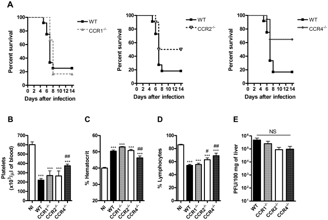
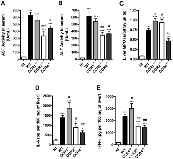
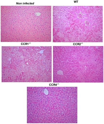
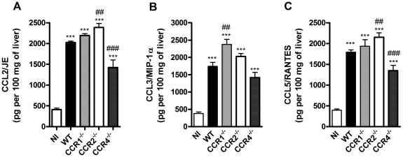
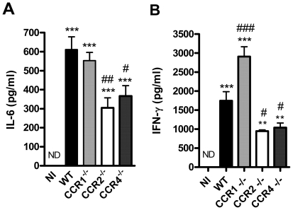
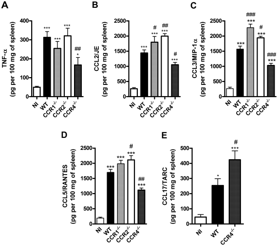
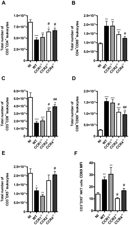
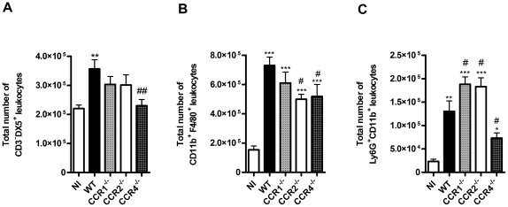
Dengue virus (DENV), a mosquito-borne flavivirus, is a public health problem in many tropical countries. Recent clinical data have shown an association between levels of different chemokines in plasma and severity of dengue. We evaluated the role of CC chemokine receptors CCR1, CCR2 and CCR4 in an experimental model of DENV-2 infection in mice. Infection of mice induced evident clinical disease and tissue damage, including thrombocytopenia, hemoconcentration, lymphopenia, increased levels of transaminases and pro-inflammatory cytokines, and lethality in WT mice. Importantly, infected WT mice presented increased levels of chemokines CCL2/JE, CCL3/MIP-1α and CCL5/RANTES in spleen and liver. CCR1⁻/⁻ mice had a mild phenotype with disease presentation and lethality similar to those of WT mice. In CCR2⁻/⁻ mice, lethality, liver damage, levels of IL-6 and IFN-γ, and leukocyte activation were attenuated. However, thrombocytopenia, hemoconcentration and systemic TNF-α levels were similar to infected WT mice. Infection enhanced levels of CCL17/TARC, a CCR4 ligand. In CCR4⁻/⁻ mice, lethality, tissue injury and systemic inflammation were markedly decreased. Despite differences in disease presentation in CCR-deficient mice, there was no significant difference in viral load. In conclusion, activation of chemokine receptors has discrete roles in the pathogenesis of dengue infection. These studies suggest that the chemokine storm that follows severe primary dengue infection associates mostly to development of disease rather than protection.


Conflict of interest statement

Competing Interests: The authors have declared that no competing interests exist.

Figures

Figure 1
Figure 1. Lethality rate, hematological alterations and viral load upon DENV-2 infection.
WT or CCR1–/–, CCR2–/– and CCR4–/– (KO) mice were infected i.p. with 10 LD50 of DENV-2 and then monitored for lethality until day 14. In panel A, percentages of survival (n = 10–12). Hematological analysis were done at day 6 p.i. for changes in platelets count (B) hematocrit (C) and lymphocytes (D) in the blood of non-infected and infected-WT and KO mice. In panel E, viral loads recovered from the liver of WT and KO mice at day 6 p.i. shown as the log of PFU/100 mg of tissue. Results are expressed as mean ± SEM and are representative of at least two experiments (n = 5–12 mice). *** P<0.001 when compared to non-infected mice. # P<0.05, ## P<0.01 when compared to WT infected mice. NI: non-infected mice. NS: Not significant.
Figure 2
Figure 2. Liver inflammation and injury upon DENV-2 infection.
WT or KO mice were infected i.p. with 10 LD50 of DENV-2 and then sacrificed at day 6 for blood and tissue samples. AST (A) and ALT (B) were dosed in serum of WT and KO mice as markers of hepatic injury. MPO activity, as an index of neutrophil accumulation, was evaluated in liver (C). Concentrations of cytokines IL-6 (D) and IFN-γ (E) were evaluated in liver homogenates by ELISA and are expressed as pg per 100 mg of tissue. Results are expressed as mean ± SEM and are representative of at least two experiments (n = 5–6). * P<0.05, *** P<0.001 when compared to non-infected mice. # P<0.05, ## P<0.01, ### P<0.001 when compared to WT infected mice. NI: non-infected mice.
Figure 3
Figure 3. Histological changes in liver upon DENV-2 infection in mice.
WT or KO mice were infected i.p. with 10 LD50 of DENV-2 and then sacrificed at day 6 for tissue samples. Hematoxylin & Eosin stained liver sections from non-infected and DENV-2 infected WT, CCR1–/–, CCR2–/– and CCR4–/– mice, showing different degrees of congestion, hemorrhage, hepatocyte degeneration and necrosis. Each slide presented in the panel is representative of at least 10 different fields (n = 5–6 mice). Magnification: 400X.
Figure 4
Figure 4. Chemokine production in liver upon DENV-2 infection in mice.
WT or KO mice were infected i.p. with 10LD50 of DENV-2 and then sacrificed at day 6 for tissue samples. CCL2/JE (A), CCL3/MIP-1α (B) and CCL5/RANTES (C) were evaluated in liver homogenates by ELISA and are expressed as pg per 100 mg of tissue. Results are expressed as mean ± SEM and are representative of at least two experiments (n = 5–6 mice). *P<0.05, *** P<0.001 when compared to non-infected mice. # P<0.05, ## P<0.01, ### P<0.001 when compared to WT infected mice. NI: non-infected mice.
Figure 5
Figure 5. Cytokine production in serum upon DENV-2 infection in mice.
WT or KO mice were infected i.p. with 10 LD50 of DENV-2 and then sacrificed at day 6 for blood samples. IL-6 (A) and IFN-γ (B) were evaluated in serum by ELISA and are expressed as pg/ml. Results are expressed as mean ± SEM and are representative of at least two experiments (n = 5–6 mice). ** P<0.01, *** P<0.001 when compared to non-infected mice. # P<0.05, ## P<0.01, ### P<0.001 when compared to WT infected mice. NI: non-infected mice.
Figure 6
Figure 6. Cytokine and chemokine production in spleen upon DENV-2 infection in mice.
WT or KO mice were infected i.p. with 10 LD50 of DENV-2 and then sacrificed at day 6 for tissue samples. TNF-α (A), CCL2/JE (B), CCL3/MIP-1α (C), CCL5/RANTES (D) and CCL17/TARC (E) were evaluated in spleen homogenates by ELISA and are expressed as pg per 100 mg of tissue. Results are expressed as mean ± SEM and are representative of at least two experiments (n = 5–6 mice). * P<0.05, *** P<0.001 when compared to non-infected mice. # P<0.05, ## P<0.01, ### P<0.001 when compared to WT infected mice. NI: non-infected mice.
Figure 7
Figure 7. Lymphocyte number and activation in CC chemokine receptors deficient mice upon DENV-2 infection.
WT or KO mice were infected i.p. with 10 LD50 of DENV-2 and then sacrificed at day 6. Splenic leukocytes were counted and then stained with specific antibodies. Flow cytometry, according to size and granularity, were performed as analysis. The numbers of specific cell populations are shown compared to total number of leukocytes in the spleen. Number of T lymphocytes CD3+CD4+ (A) and CD3+CD8+ (C) were evaluated in WT and KO mice. Activated T lymphocytes expressing CD69 were also evaluated for CD4+ (B) and CD8+ (D) populations. The number of CD3+DX5+ NKT cells (E) and their activation by CD69 expression as MFI, were also evaluated (F). Results are expressed as mean ± SEM and are representative of at least two experiments (n = 5–6 mice). *P<0.05, ** P<0.01, *** P<0.001 when compared to non-infected mice. # P<0.05, ## P<0.01 when compared to WT infected mice. NI: non-infected mice. MFI: Mean fluorescence intensity.
Figure 8
Figure 8. NK, macrophages and neutrophils number in CC chemokine receptors deficient mice upon DENV-2 infection.
WT or KO mice were infected i.p. with 10 LD50 of DENV-2 and then sacrificed at day 6. Splenic leukocytes were counted and then stained with specific antibodies. Flow cytometry, according to size and granularity, were performed as analysis. The numbers of specific cell populations are shown compared to total number of leukocytes in the spleen. Numbers of CD3DX5+ NK cells (A), macrophages CD11b+F4/80+ (B) and infiltrating neutrophils CD11b+Ly6G+ (C) were evaluated. Results are expressed as mean ± SEM and are representative of at least two experiments (n = 5–6 mice). *P<0.05, ** P<0.01, *** P<0.001 when compared to non-infected mice. # P<0.05, ## P<0.01 when compared to WT infected mice. NI: non-infected mice.

Similar articles

Cited by

References

    1. Guzman MG, Kouri G. Dengue: an update. Lancet Infect Dis. 2002;2:33–42. - PubMed
    1. Mackenzie JS, Gubler DJ, Petersen LR. Emerging flaviviruses: the spread and resurgence of Japanese encephalitis, West Nile and dengue viruses. Nat Med. 2004;10:S98–109. - PubMed
    1. Kalayanarooj S, Vaughn DW, Nimmannitya S, Green S, Suntayakorn S, et al. Early clinical and laboratory indicators of acute dengue illness. J Infect Dis. 1997;176:313–321. - PubMed
    1. Deen JL, Harris E, Wills B, Balmaseda A, Hammond SN, et al. The WHO dengue classification and case definitions: time for a reassessment. Lancet. 2006;368:170–173. - PubMed
    1. Gibbons RV, Vaughn DW. Dengue: an escalating problem. BMJ. 2002;324:1563–1566. - PMC - PubMed

Publication types