. 2011 Apr;60(4):525-35.
doi: 10.1007/s00262-010-0961-7. Epub 2011 Jan 5.

Deficiency of activated STAT1 in head and neck cancer cells mediates TAP1-dependent escape from cytotoxic T lymphocytes

Affiliations

Deficiency of activated STAT1 in head and neck cancer cells mediates TAP1-dependent escape from cytotoxic T lymphocytes

Michael S Leibowitz et al. Cancer Immunol Immunother. 2011 Apr.

Abstract

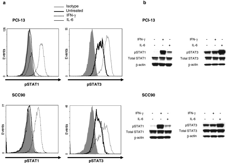
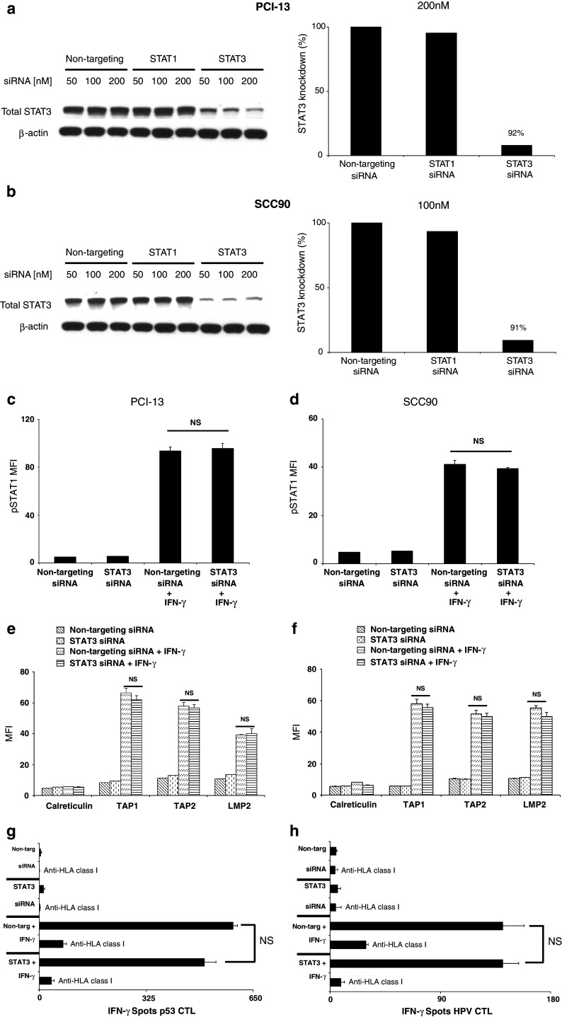
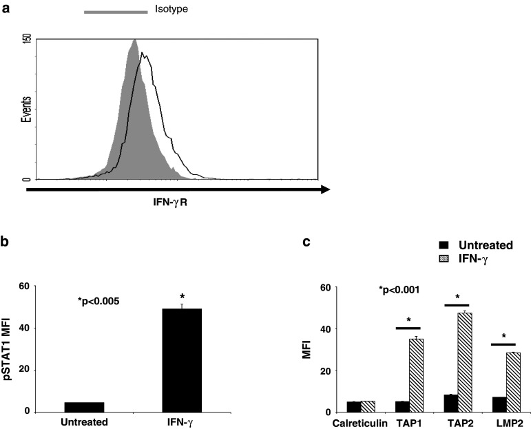
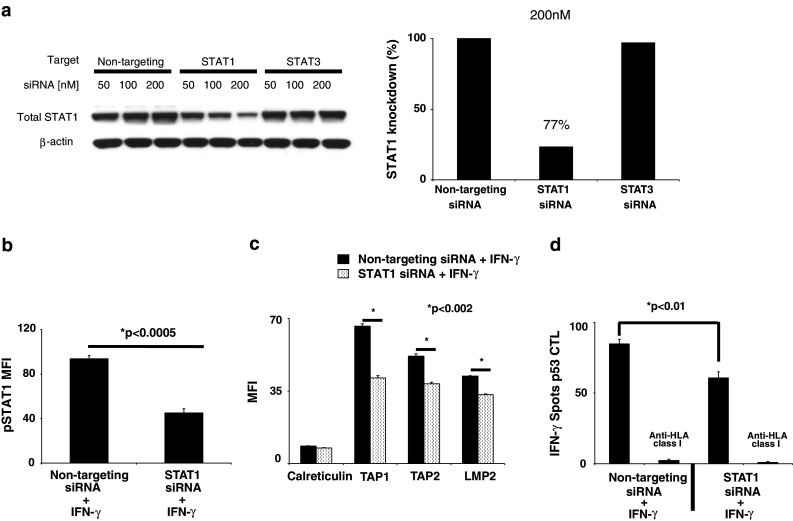
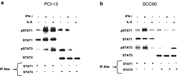
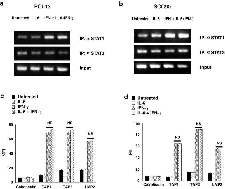
Squamous cell carcinoma of the head and neck (SCCHN) cells can escape recognition by tumor antigen (TA)-specific cytotoxic T lymphocytes (CTL) by downregulation of antigen processing machinery (APM) components, such as the transporter associated with antigen processing (TAP)-1/2 heterodimer. APM component upregulation by interferon gamma (IFN-γ) restores SCCHN cell recognition and susceptibility to lysis by CTL, but the mechanism underlying TAP1/2 downregulation in SCCHN cells is not known. Because IFN-γ activates signal transducer and activator of transcription (STAT)-1, we investigated phosphorylated (p)-STAT1 as a mediator of low basal TAP1/2 expression in SCCHN cells. SCCHN cells were found to express basal total STAT1 but low to undetectable levels of activated STAT1. The association of increased pSTAT1 levels and APM components likely reflects a cause-effect relationship, since STAT1 knockdown significantly reduced both IFN-γ-mediated APM component expression and TA-specific CTL recognition of IFN-γ-treated SCCHN cells. On the other hand, since oncogenic pSTAT3 is overexpressed in SCCHN cells and was found to heterodimerize with pSTAT1, we also tested whether pSTAT3 and pSTAT1:pSTAT3 heterodimers inhibited IFN-γ-induced STAT1 activation and APM component expression. First, STAT3 activation or depletion did not affect basal or IFN-γ-induced expression of pSTAT1 and APM components or recognition of SCCHN cells by TA-specific CTL. Second, pSTAT1:pSTAT3 heterodimers did not interfere with IFN-γ-induced STAT1 binding to the TAP1 promoter or APM protein expression. These findings demonstrate that APM component downregulation is regulated primarily by an IFN-γ-pSTAT1-mediated signaling pathway, independent of oncogenic STAT3 overexpression in SCCHN cells.


Figures

Fig. 1
Fig. 1
SCCHN cells express low basal pSTAT1 and high basal pSTAT3 that are inducible by treatment with IFN-γ and IL-6. Cells were treated with IFN-γ (100 U/ml, 15 min) or IL-6 (50 ng/ml, 15 min) and assayed for pSTAT1 (Tyrosine 701) or pSTAT3 (Tyrosine 705) by a intracellular flow cytometry or b immunoblot analyses. Data represent at least three independent experiments
Fig. 2
Fig. 2
IFN-γ-mediated pSTAT1, APM expression and CTL recognition in STAT3-depleted SCCHN cells. a PCI-13 and b SCC90 cells were transfected with the indicated doses of siRNA, and knockdown of STAT3 protein was assessed by immunoblot and densitometry analyses. Data represent at least three independent experiments. c PCI-13 and d SCC90 cells were transfected with (200 or 100 nM, respectively) non-targeting or STAT3 siRNA alone or in combination with IFN-γ (100 U/ml, 15 min), added 48 h after transfection. Intracellular flow cytometry for pSTAT1 was performed. Data represent the mean fluorescence intensity MFI of at least three independent experiments. (P = NS, two-tailed t test). Error bars indicate standard error. PCI-13 (e) and SCC90 (f) cells were transfected as described above, except IFN-γ (100 U/ml, 48 h) was added 24 h after transfection. Intracellular flow cytometry for TAP1, TAP2, LMP2 and calreticulin was performed. Calreticulin, a non-IFN-γ-inducible APM component, was included to control for global changes in protein expression following treatment. Data represent the (MFI) of at least three independent experiments. (P = NS, two-tailed t test). Error bars indicate standard error. IFN-γ ELISPOT assay were performed using (g) p5365–73- or (h) HPV7–15 -specific cytotoxic T lymphocytes as effector cells and SCC90 cells as targets that have been transfected and treated with IFN-γ as described in (b). An anti-HLA-A,B,C mAb (w6/32) was used to demonstrate that CTL was HLA class I restricted. Error bars indicate standard error (P = NS, two-tailed t test)
Fig. 3
Fig. 3
STAT1 signaling is intact in SCCHN cells. a SCCHN cells isolated from SCCHN tumors were tested for basal IFN-γR expression by flow cytometry. PCI-13 cells were treated with IFN-γ (100 U/ml) for either 15 min or 48 h and then analyzed by intracellular flow cytometry for either b pSTAT1 c or APM component expression, respectively. Data represent at least three independent experiments. Mean fluorescence intensity (MFI) was plotted, and error bars indicate standard error (*P < 0.005, *P < 0.001, two-tailed t test)
Fig. 4
Fig. 4
Activated STAT1 is a critical mediator of APM component expression and TA-specific CTL recognition of SCCHN cells. a PCI-13 cells were transfected with the indicated doses of siRNA, and knockdown of STAT1 protein was assessed by immunoblot and densitometry analyses. PCI-13 cells were transfected with 200 nM of the indicated siRNA and 24 h after transfection, IFN-γ (40 U/ml) was added for either 15 min or 48 h and then analyzed by intracellular flow cytometry for either b pSTAT1 or c APM component expression by MFI, respectively. Data represent at least three independent experiments. Error bars indicate standard error (*P < 0.0005, *P < 0.002, two-tailed t test). d PCI-13 cells were transfected with 200 nM of the indicated siRNA and 24 h after transfection treated with IFN-γ (40 U/ml) for an additional 24 h. The cells were collected as used as targets, and p5365–73-specific cytotoxic T lymphocyte (CTL) was used as effector cells in IFN-γ ELISPOT assays. An anti-HLA-A,B,C mAb (w6/32) was used to demonstrate that CTL was HLA class I restricted. Error bars indicate standard error (*P < 0.01, two-tailed t test)
Fig. 5
Fig. 5
IL-6 and IFN-γ increase pSTAT1:pSTAT3 heterodimerization. Cells were treated with either IL-6 (50 ng/ml, 15 min) or IFN-γ (100 U/ml, 15 min). Whole cell lysates were prepared and immunoprecipitated with anti-STAT1 or anti-STAT3 or an irrelevant control mAb. The immunoprecipitates were size fractionated by SDS/PAGE and transferred to a PVDF membrane. The blots were probed for anti-pSTAT1, STAT1, pSTAT3, and total STAT3 from a PCI-13 and b SCC90 cells
Fig. 6
Fig. 6
IFN-γ-mediated-STAT1 binding to the TAP1 promoter and APM protein expression is independent of STAT1:STAT3 heterodimerization. a PCI-13 and b SCC90 cells were untreated, treated with IL-6 (50 ng/ml, 60 min), IFN-γ (1,000 U/ml, 30 min) or pretreated with IL-6 (50 ng/ml, 30 min), then treated with IFN-γ (1,000 U/ml, 30 additional min) in the presence of IL-6. The cells were fixed with formaldehyde, quenched with glycine and lysed in SDS lysis buffer. Chromatin was sheared by sonication and probed with anti-STAT1, anti-STAT3 and anti-IgG mAbs. Protein–DNA crosslinks were reversed, and both RNA and protein were removed by enzymatic digestion. DNA was purified, and PCR was performed amplifying a canonical GAS sequence in the TAP1 promoter localized to STAT1 binding. c PCI-13 and d SCC90 cell were untreated, treated with IL-6 (50 ng/ml, 48 h), IFN-γ (100 U/ml, 48 h) or pretreated with IL-6 (50 ng/ml, 30 min), then treated with IFN-γ (100 U/ml, 48 h) in the presence of IL-6. Intracellular flow cytometry was performed measuring TAP1, TAP2, LMP2 and calreticulin. MFI was plotted representing at least three independent experiments (P = NS, two-tailed t test). Error bars indicate standard error

Similar articles

Cited by

References

    1. Cerundolo V, et al. Presentation of viral antigen controlled by a gene in the major histocompatibility complex. Nature. 1990;345(6274):449–452. doi: 10.1038/345449a0. - DOI - PubMed
    1. Hosken NA, Bevan MJ. Defective presentation of endogenous antigen by a cell line expressing class I molecules. Science. 1990;248(4953):367–370. doi: 10.1126/science.2326647. - DOI - PubMed
    1. Campoli M, Ferrone S. HLA antigen changes in malignant cells: epigenetic mechanisms and biologic significance. Oncogene. 2008;27(45):5869–5885. doi: 10.1038/onc.2008.273. - DOI - PMC - PubMed
    1. Seliger B. Different regulation of MHC class I antigen processing components in human tumors. J Immunotoxicol. 2008;5(4):361–367. doi: 10.1080/15476910802482870. - DOI - PubMed
    1. Ferris RL, Hunt JL, Ferrone S. Human leukocyte antigen (HLA) class I defects in head and neck cancer: molecular mechanisms and clinical significance. Immunol Res. 2005;33(2):113–133. doi: 10.1385/IR:33:2:113. - DOI - PubMed

MeSH terms

Substances