. 2011 Sep;68(18):3081-93.
doi: 10.1007/s00018-010-0608-z. Epub 2011 Jan 5.

GSK-3β is essential for physiological electric field-directed Golgi polarization and optimal electrotaxis

Affiliations

GSK-3β is essential for physiological electric field-directed Golgi polarization and optimal electrotaxis

Lin Cao et al. Cell Mol Life Sci. 2011 Sep.

Abstract

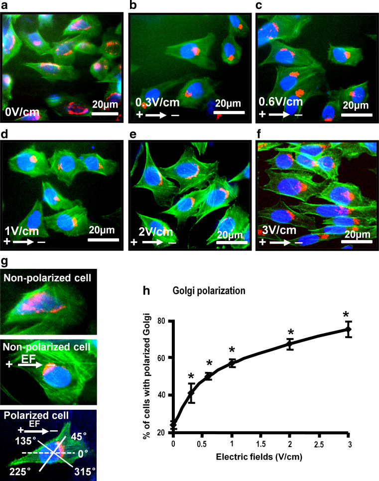
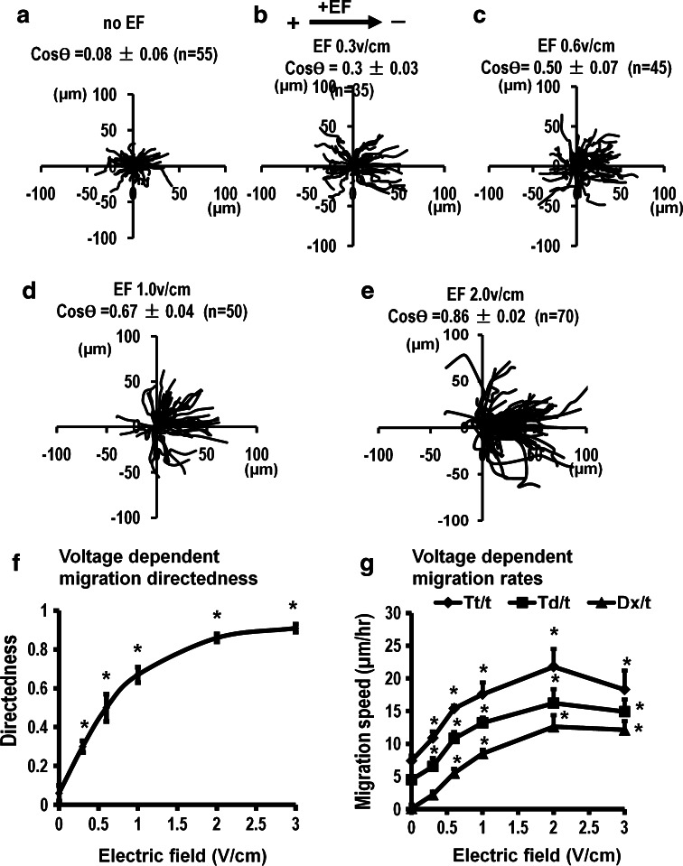
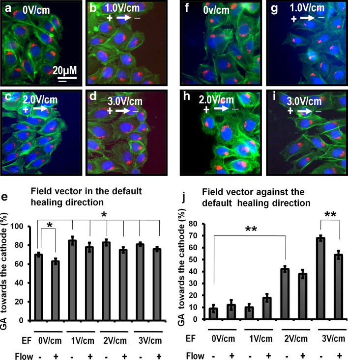
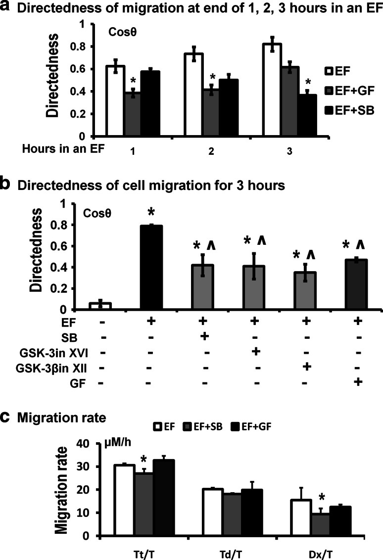
Endogenous electrical fields (EFs) at corneal and skin wounds send a powerful signal that directs cell migration during wound healing. This signal therefore may serve as a fundamental regulator directing cell polarization and migration. Very little is known of the intracellular and molecular mechanisms that mediate EF-induced cell polarization and migration. Here, we report that Chinese hamster ovary (CHO) cells show robust directional polarization and migration in a physiological EF (0.3-1 V/cm) in both dissociated cell culture and monolayer culture. An EF of 0.6 V/cm completely abolished cell migration into wounds in monolayer culture. An EF of higher strength (≥1 V/cm) is an overriding guidance cue for cell migration. Application of EF induced quick phosphorylation of glycogen synthase kinase 3β (GSK-3β) which reached a peak as early as 3 min in an EF. Inhibition of protein kinase C (PKC) significantly reduced EF-induced directedness of cell migration initially (in 1-2 h). Inhibition of GSK-3β completely abolished EF-induced GA polarization and significantly inhibited the directional cell migration, but at a later time (2-3 h in an EF). Those results suggest that GSK-3β is essential for physiological EF-induced Golgi apparatus (GA) polarization and optimal electrotactic cell migration.


Figures

Fig. 1
Fig. 1
EFs of physiological strength directed GA polarization in CHO cells. af Fluorescent images showed GA (red) in CHO cells cultured in the absence (a) or presence of an EF for 3 h. The cells were fixed and triple-labeled with GM130 antibody (GA marker, red), FITC-phalloidin (F-actin, green), and DAPI (blue). g GA polarization was quantified as the percentage of GA polarized into the quadrant between 45 and 315 degrees in the field direction as shown. h An EF of 0.3 V/cm induced significant GA polarization, which was more pronounced with increased field strength. *P < 0.01 compared to no EFs control. Scale bar 20 μm
Fig. 2
Fig. 2
EFs of physiological strength directed migration of CHO cells. ae The migration trajectories of CHO cells for a period of 3 h in absence or presence of an EF showed significant directional migration in an EF of 0.3 V/cm or higher voltage. Each cell’s position at 0 h is positioned at the origin (0, 0). f Voltage dependence of migration directedness of CHO cells showed a threshold below 0.5 V/cm. g Trajectory speed and displacement speed along the x-axis all increased with the field strength of the applied EFs. All results were from three independent experiments. *P < 0.01 compared to no field control. Directedness and migration speed values are shown as mean ± SEM, with total number of cells as n
Fig. 3
Fig. 3
A physiological EF directed GA polarization in the scratch wound model. ad An EF applied in the default healing direction significantly increased GA polarization into the wound. e GA polarization in the first rows of cells at wound edge was shown. EFs of 1.0, 2.0 and 3.0 V/cm in the default healing direction significantly increased GA polarization into the wound to the same extent. GA polarization at 1.0, 2.0 and 3.0 V/cm was reduced by a continuous medium flow perpendicular to the EF lines with no significant difference. fi An EF applied against the default healing direction polarized the GA away from the wound. j GA polarization in the first row at EF against wound edge with/without medium flow was shown. GA polarization at 3.0 V/cm was significantly reduced in medium flow. The medium flow was maintained during the experiments to exclude possible chemotaxis. The flow of copious culture medium through the electrotactic chamber was maintained with a peristaltic pump during the entire experimental period (24 mL/h, through 0.2 mm2 channel). Data were grouped from three independent experiments. The percentage of cells with GA polarized in the EF direction is shown as mean ± SEM. Monolayer culture of CHO cells were wounded and allowed to heal for 3 h (ai). The cells were triple-labeled with actin (green), GM130 (red), and DAPI (blue). *P < 0.05, **P < 0.01 when compared to control. Bar 20 μm
Fig. 4
Fig. 4
A physiological EF was a predominant cue directing cell migration in monolayer wound healing. a Cells at one side of the wound edges migrated to the left. The following experiments were done on this side of the wound. b Migration trajectories of cells at the wound in the absence of an EF were shown. cf An EF was applied with the field vector against the normal healing direction. Each cell’s position at t = 0 is positioned at the origin. Trajectories of cell migration were shown for a period 3 h in the EF. Migration of the cells into the wound was significantly reduced at 0.3 V/cm, and was stopped at 0.6 V/cm. The migration direction was completely reversed and cells migrated away from the wound in an EF of 1 and 2 V/cm. g Directedness of cell migration was voltage dependent with a threshold less than 1 V/cm. Cos θ is mean values of directedness ± SEM. n the total number of cells at the given condition
Fig. 5
Fig. 5
An EF induced the phosphorylation of GSK-3β. a, b (upper channels) Time courses of phosphorylation of Akt and GSK-3β were analyzed with western blotting. a, b (lower channels) The expression of total GSK-3β and Akt were no different between the EF-supplied and no EF control. c GF109203X (GF, PKC inhibitor) completely inhibited the phosphorylation of GSK-3β. d SB216763 (SB, inhibitor of GSK-3β) did not inhibit the phosphorylation of GSK-3β. e Phosphorylation of GSK-3β could be inhibited by inhibitors of PI3K (30 μM Ly294002 and 200 nM Wortmannin) and inhibitor of PKC (20 μM GF109203X). GF109203X (GF, PKC inhibitor) completely inhibited the phosphorylation of GSK-3β. f β-catenin as a marker of GSK-3 function was down-regulated by EF. Inhibition of GSK-3 with 1 μM GSK-3 inhibitor XVI (GSK-3in XVI) or GSK-3β with 1 μM GSK-3β inhibitor XII (GSK-3βin XII) could increase the expression of β-catenin. α-Tubulin or GAPDH was used as protein loading control. Histograms show the relative intensity of phospho-GSK3β and phospho-Akt over the tubulin control. EF = 2.0 V/cm. All results were from three independent experiments
Fig. 6
Fig. 6
GSK-3β was essential for EF-directed GA polarization. a, b 20 μM GF109203X (PKC inhibitor) could partially reduce the EF-induced GA polarization. However, inhibitions of GSK-3 or GSK-3β with 20 μM SB216763, 1 μM GSK-3 inhibitor XVI (GSK-3in XVI) or 1 μM GSK-3β inhibitor XII (GSK-3β in XII), could abolish the GA polarization induced by EF. CHO cells were pre-treated with GSK-3β inhibitor, 20 μM SB216763, 1 μM GSK-3 inhibitor XVI (GSK-3in XVI), 1 μM GSK-3β inhibitor XII (GSK-3β in XII) or 20 μM GF109203X (PKC inhibitor) for 1 h and then subjected to 1.5–2.0 V/cm of EFs for 3 h. The cells were labeled with GM130 (red) and DAPI (blue). *P < 0.05 when compared with the no field control. ^ P < 0.05 compared the field no drug control. Values are shown as mean ± SEM. All results were from three independent experiments
Fig. 7
Fig. 7
GA polarization partially contributes the EF-directed cell migration. CHO cells were pre-treated with GSK-3β inhibitor, 20 μM SB216763 (SB), 1 μM GSK-3 inhibitorXVI (GSK-3in XVI), 1 μM GSK-3β inhibitor XII (GSK-3β in XII) or 20 μM GF109203X (GF, PKC inhibitor), and then applied to 1.5–2.0 V/cm EF for 3 h. a Migration directedness at the end of the first, second and third hour. The inhibitory effect of GF was significant 1 h in the EF, while the inhibition of SB appeared at the end of 3 h in an EF. b Inhibition of GSK-3, GSK-3β or PKC significantly inhibited the directedness of EF-directed migration of CHO cells. c The migration speed was inhibited when cell to be treated with inhibitors of GSK-3β. But the inhibitors of PKC did not reduce the migration speed. All results were from three independent experiments. *P < 0.01 compared with no EF control. ^P < 0.01 compared with EF control. Values are shown as mean ± SEM, from three independent experiments with total cell numbers of 50

Similar articles

Cited by

References

    1. Jaffe LF, Vanable JW., Jr Electric fields and wound healing. Clin Dermatol. 1984;2:34–44. doi: 10.1016/0738-081X(84)90025-7. - DOI - PubMed
    1. McCaig CD, Zhao M. Physiological electrical fields modify cell behaviour. BioEssays. 1997;19:819–826. doi: 10.1002/bies.950190912. - DOI - PubMed
    1. McCaig CD, Rajnicek AM, Song B, Zhao M. Has electrical growth cone guidance found its potential? Trends Neurosci. 2002;25:354–359. doi: 10.1016/S0166-2236(02)02174-4. - DOI - PubMed
    1. Nuccitelli R. A role for endogenous electric fields in wound healing. Curr Top Dev Biol. 2003;58:1–26. doi: 10.1016/S0070-2153(03)58001-2. - DOI - PubMed
    1. Nuccitelli R. Endogenous electric fields in embryos during development, regeneration and wound healing. Radiat Prot Dosimetry. 2003;106:375–383. - PubMed

Publication types

Substances