. 2011 May-Jun;87(3):699-706.
doi: 10.1111/j.1751-1097.2011.00890.x. Epub 2011 Feb 10.

Effects of endosomal photodamage on membrane recycling and endocytosis

Affiliations

Effects of endosomal photodamage on membrane recycling and endocytosis

Michelle Andrzejak et al. Photochem Photobiol. 2011 May-Jun.

Abstract

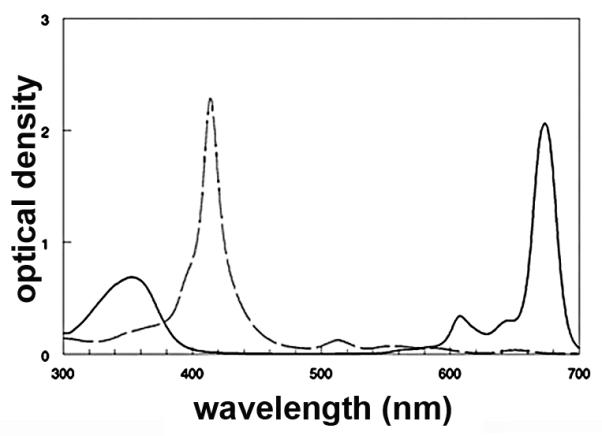
The flux of receptor-independent endocytosis can be estimated by addition of wortmannin to cell cultures. Membrane influx is unaffected but traffic out of late endosomes is impaired, resulting in a substantial enlargement of these organelles. Using the 1c1c7 murine hepatoma, we investigated the effect of endosomal photodamage on this endocytic pathway. We previously reported that photodamage catalyzed by the lysosomal photosensitizer NPe6 prevented wortmannin-induced endosomal swelling, indicating an earlier block in the process. In this study, we show that endosomal photodamage, initiated by photodamage from an asymmetrically substituted porphine or a phthalocyanine also prevents wortmannin-induced endosomal swelling, even when the photodynamic therapy (PDT) dose is insufficient to cause endosomal disruption. As the PDT dose is increased, endosomal breakage occurs, as does apoptosis and cell death. Very high PDT doses result in necrosis. We propose that photodamage to endosomes results in alterations in the endosomal structure such that influx of new material is inhibited and receptor-independent endocytosis is prevented. In an additional series of studies, we found that the swollen late endosomes induced by wortmannin are unable to retain previously accumulated fluorescent probes or photosensitizers.


Figures

Figure 1
Figure 1
Absorbance spectra of 10 μM AlPcS2a (solid line) and TPPS2a (dotted line) in ethanol.
Figure 2
Figure 2
Production of swollen late endosomes 30 min after treatment of 1c1c7 cells with 20 nM wortmannin. A, no additional treatment; B-E, after irradiation of cells (670 ± 10 nm) previously loaded with 5 μM AlPCS2a as described in the text: B = 15 mJ/cm2; C = 30 mJ/cm2, D = 50 mJ/cm2 , E = 140 mJ/cm2 of light at 670 ±10 nm. White bar in panel A = 20 μm.
Figure 3
Figure 3
Phase-contrast and fluorescent images of cells loaded with 5 μM AlPcS2a as described in the text then irradiated at 670 ± 10 nm with varying light doses (mJ/cm2): A,B = 0; C,D =15; E,F = 30; G,H = 55; I,J = 140. White bar in panel A = 20 μm.
Figure 4
Figure 4
Phase contrast (left) and fluorescence (right) images of 1c1c7 cells loaded with 5 μM AlPcS2a as described in the text, then irradiated (670 ± 10 nm) with 0 (A), 55 (B), 140 (C) or 270 (D) mJ/cm2. Cells were subsequently incubated for 60 min at 37°;C with HO342 added for the last 10 min of these incubations. White bar in panel A = 50 μm. Arrows in panels C and D indicate apoptotic cells; in panel G, swollen pre-necrotic cells.
Figure 5
Figure 5
Localization of TPPS2a and AlPcS2a. Left panels: phase contrast images; right panels, fluorescence. A,B = TPPS2a after a 1 hr incubation. C,D = TPPS2a uptake directly following photodamage induced by NPe6 (LD30). E,F = AlPcS2a uptake after 1 hr. G,H = AlPcS2a uptake following NPe6-induced photodamage. White bar in panel A = 20 μm.
Figure 6
Figure 6
Labeling of 1c1c7 cells by TDPH. Left panels, phase contrast; right panels, fluorescence. A = TDPH 30 sec incubation B = TDPH 30 sec incubation a 30 min incubation in fresh medium at 37°;C. C, Similar to B using cells loaded with 5 μM AlPS2a and irradiated (30 mJ/cm2). White bar in panel A = 20 μm.
Figure 7
Figure 7
Cells incubated with 5 μM AlPcS2a for 16 hr, then washed for 1 h with 20 nM wortmannin added during the final 30 min. A = phase contrast; B = fluorescence. Panel C shows the effect of a 30 mJ/cm2 light dose before addition of wortmannin. White bar in panel A = 20 μm.
Figure 8
Figure 8
Release of LSG by wortmannin. Cells were incubated with 1 μM LSG for 10 min, then placed in fresh medium for 30 min at 37°;C (A), or in medium containing 20 nM wortmannin for 30 min (B). White bar in panel A = 20 μm.

References

    1. Dougherty TJ, Gomer CJ, Henderson BW, Jori G, Kessel D, Korbelik M, Moan J, Peng Q. Photodynamic therapy. J. Natl. Cancer Inst. 1998;90:889–905. - PMC - PubMed
    1. Kessel D, Oleinick NL. Photodynamic therapy and cell death pathways. Methods Mol. Biol. 2010;635:35–46. - PMC - PubMed
    1. Reiners JJ, Jr, Agostinis P, Berg K, Oleinick NL, Kessel D. Assessing autophagy in the context of photodynamic therapy. Autophagy. 2010;6:7–18. - PMC - PubMed
    1. Ouédraogo G, Morlière P, Mazière C, Mazière JC, Santus R. Alteration of the endocytotic pathway by photosensitization with fluoroquinolones. Photochem. Photobiol. 2000;72:458–463. - PubMed
    1. Olchawa M, Szewczyk G, Zareba M, Pilat A, Bzowska M, Mikolajczyk T, Sarna T. Sub-lethal Photodynamic Damage to ARPE-19 Cells Transiently Inhibits Their Phagocytic Activity. Photochem. Photobiol. 2010;86:772–780. - PubMed

Publication types

MeSH terms