. 2011 Mar 11;286(10):8188-8196.
doi: 10.1074/jbc.M110.192013. Epub 2011 Jan 5.

A compact beta model of huntingtin toxicity

Affiliations

A compact beta model of huntingtin toxicity

Qi Charles Zhang et al. J Biol Chem. .

Abstract

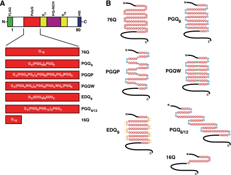
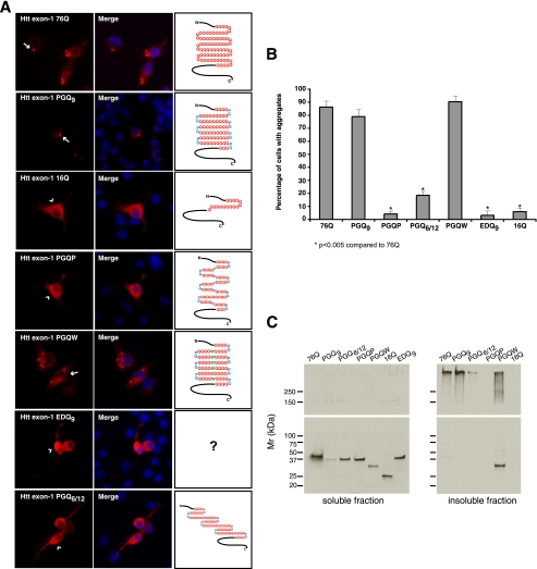
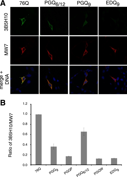
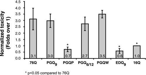
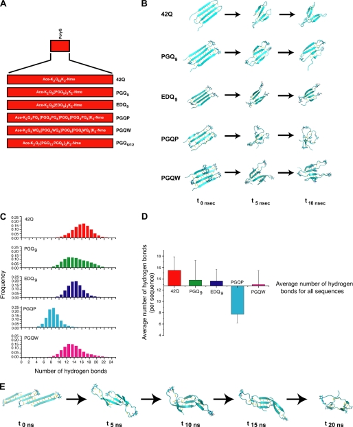
Huntington disease results from an expanded polyglutamine region in the N terminus of the huntingtin protein. HD pathology is characterized by neuronal degeneration and protein inclusions containing N-terminal fragments of mutant huntingtin. Structural information is minimal, though it is believed that mutant huntingtin polyglutamine adopts β structure upon conversion to a toxic form. To this end, we designed mammalian cell expression constructs encoding compact β variants of Htt exon 1 N-terminal fragment and tested their ability to aggregate and induce toxicity in cultured neuronal cells. In parallel, we performed molecular dynamics simulations, which indicate that constructs with expanded polyglutamine β-strands are stabilized by main-chain hydrogen bonding. Finally, we found a correlation between the reactivity to 3B5H10, an expanded polyglutamine antibody that recognizes a compact β rich hairpin structure, and the ability to induce cell toxicity. These data are consistent with an important role for a compact β structure in mutant huntingtin-induced cell toxicity.


Figures

FIGURE 1.
FIGURE 1.
Htt exon-1 polyQ and compact β polyQ proteins used for mammalian cell culture transfection experiments. A, domain structure of Htt exon-1 fragment, indicating polyQ region (red). Also shown are two Pro-repeat regions (yellow) and the P/Q-rich region (purple). An N-terminal FLAG tag (green) and a C-terminal His6 tag (blue) were engineered into each construct. The primary structure for each polyQ region is shown. B, predicted secondary structure of polyQ region in the expressed protein.
FIGURE 2.
FIGURE 2.
Aggregation propensities of Htt exon-1 polyQ and compact β polyQ proteins in transiently transfected N2a cells. A, immunofluorescence images of transfected N2a cells showing MW7 staining (red, left panels) 48 h after transfection. Arrows indicate Htt exon-1 76Q, PGQ9, and PGQW cytoplasmic and perinuclear aggregates, while arrowheads show diffuse cytoplasmic staining in Htt exon-1 16Q, PGQP-, EDQ9-, and PGQ6/12-expressing cells. Nuclei in merged images were stained with Hoechst (blue, middle panels). B, quantification of aggregated Htt exon-1 by cell counting analysis. Data shown are the average (± S.D.) of three independent experiments. C, Western blot analysis of Htt exon-1 expression 48 h after transfection. Detergent-soluble supernatant (left panels) and insoluble pellet fraction (right panels) samples were prepared as described under “Experimental Procedures.” Htt exon-1 EDQ9 remained mostly soluble by IIF, as 16Q, and was not included in the insoluble fraction samples (right panels). Blots were stained with anti-FLAG (upper panels) and anti-Htt exon-1 (Ref. , lower panels) antibodies.
FIGURE 3.
FIGURE 3.
Reactivity of Htt exon-1 polyQ and compact β polyQ proteins with 3B5H10, an antibody that recognizes compact, two-stranded hairpins of polyQ, and MW7, an antibody that recognizes a wide range of Htt species, in transiently-transfected mammalian cells. A, selected immunofluorescence images of transfected HT22 cells showing 3B5H10 (green, upper panels) and MW7 (red, middle panels) staining 48 h after transfection. Nuclei in merged images were stained with Hoechst (blue, bottom panels). B, quantification of 3B5H10 reactivity, relative to MW7 staining, for Htt exon-1 polyQ and compact β polyQ proteins. All proteins are compared with Htt exon-1 76Q. Data shown are the average (± S.D.) of three independent experiments.
FIGURE 4.
FIGURE 4.
Toxicity of Htt exon-1 polyQ and compact β polyQ proteins in N2a cells. pSIVA-based suspension assay demonstrates that Htt exon-1 76Q, PGQ9, PGQ6/12, and PGQW constructs are toxic, while Htt exon-1 PGQP and EDQ9 are not toxic. Cells were stained with pSIVA, a membrane polarity-sensitive annexin-based green fluorescent probe that can be used to detect dead or dying cells (31), 72 h after transfection. Cells were then fixed and stained with MW7 antibody and processed as described under “Experimental Procedures.” Toxicity values are reported relative to non-expanded Htt exon-1 16Q, which has been previously shown in vitro and in vivo using numerous assays to be soluble and non-toxic. Data shown are the average (± S.D.) of three independent experiments.
FIGURE 5.
FIGURE 5.
Molecular dynamics simulation studies. A, schematic representation of polyQ and compact β polyQ sequences. These sequences are analogous to the constructs presented in Fig. 1A. B, snapshots for each sequence at the initial (t0 ns), midway (t5 ns), and final (t10 ns) time points of each 10 ns simulation. (C) Histograms of the number of main-chain hydrogen bonds over the 10-ns MD simulation for each sequence. D, average number of main-chain hydrogen bonds for each structure. Error bars represent root mean squared (rms) fluctuations in the number of hydrogen bonds with respect to the average. E, snapshots of PGQ6/12 at the initial (t0 ns), midway (t5 ns, t10 ns, t15 ns), and final (t20 ns) time points over the 20-ns molecular dynamics simulation.

References

    1. Ross C. A., Margolis R. L., Rosenblatt A., Ranen N. G., Becher M. W., Aylward E. (1997) Medicine 76, 305–338 - PubMed
    1. Zoghbi H. Y., Orr H. T. (2000) Annu. Rev. Neurosci. 23, 217–247 - PubMed
    1. Huntington's Disease Collaborative Research Group (1993) Cell 72, 971–983 - PubMed
    1. Ross C. A., Tabrizi S. J. (2011) Lancet 10, 83–98 - PubMed
    1. Andrade M. A., Bork P. (1995) Nat. Genet. 11, 115–116 - PubMed

Publication types

LinkOut - more resources