. 2011 Mar 4;286(9):7214-26.
doi: 10.1074/jbc.M110.169342. Epub 2011 Jan 5.

S100B protein stimulates microglia migration via RAGE-dependent up-regulation of chemokine expression and release

Affiliations

S100B protein stimulates microglia migration via RAGE-dependent up-regulation of chemokine expression and release

Roberta Bianchi et al. J Biol Chem. .

Abstract

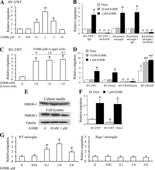
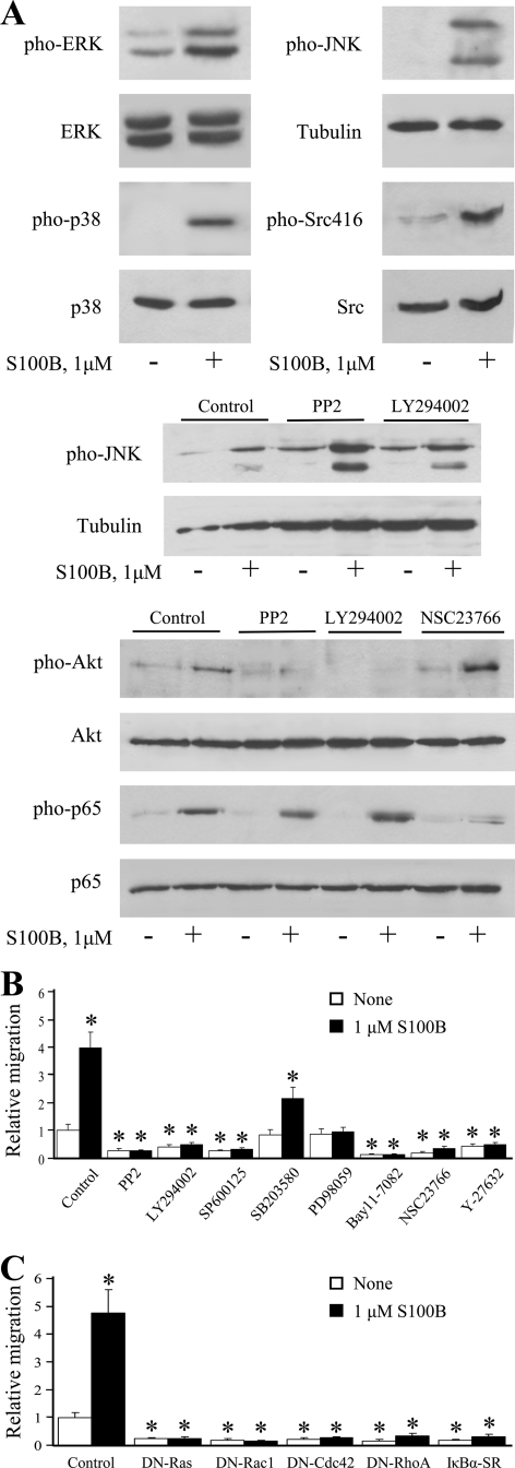
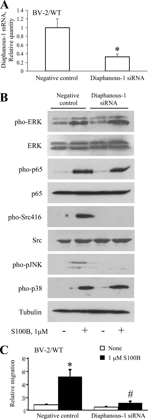
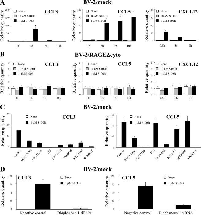
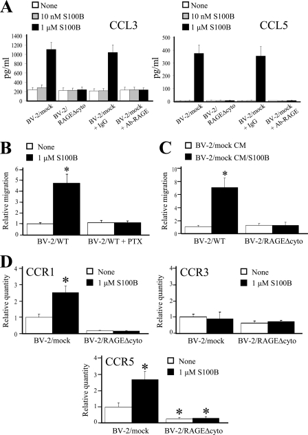
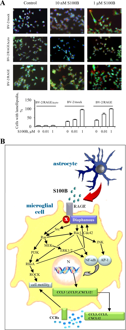
The Ca(2+)-binding protein of the EF-hand type, S100B, is abundantly expressed in and secreted by astrocytes, and release of S100B from damaged astrocytes occurs during the course of acute and chronic brain disorders. Thus, the concept has emerged that S100B might act an unconventional cytokine or a damage-associated molecular pattern protein playing a role in the pathophysiology of neurodegenerative disorders and inflammatory brain diseases. S100B proinflammatory effects require relatively high concentrations of the protein, whereas at physiological concentrations S100B exerts trophic effects on neurons. Most if not all of the extracellular (trophic and toxic) effects of S100B in the brain are mediated by the engagement of RAGE (receptor for advanced glycation end products). We show here that high S100B stimulates murine microglia migration in Boyden chambers via RAGE-dependent activation of Src kinase, Ras, PI3K, MEK/ERK1/2, RhoA/ROCK, Rac1/JNK/AP-1, Rac1/NF-κB, and, to a lesser extent, p38 MAPK. Recruitment of the adaptor protein, diaphanous-1, a member of the formin protein family, is also required for S100B/RAGE-induced migration of microglia. The S100B/RAGE-dependent activation of diaphanous-1/Rac1/JNK/AP-1, Ras/Rac1/NF-κB and Src/Ras/PI3K/RhoA/diaphanous-1 results in the up-regulation of expression of the chemokines, CCL3, CCL5, and CXCL12, whose release and activity are required for S100B to stimulate microglia migration. Lastly, RAGE engagement by S100B in microglia results in up-regulation of the chemokine receptors, CCR1 and CCR5. These results suggests that S100B might participate in the pathophysiology of brain inflammatory disorders via RAGE-dependent regulation of several inflammation-related events including activation and migration of microglia.


Figures

FIGURE 1.
FIGURE 1.
S100B stimulates BV-2 microglia migration in a RAGE-dependent manner. A, migration assays were performed using Boyden chambers. The cells (1 × 10⁵) were seeded onto the top of Transwell® migration chambers and allowed to migrate for 6 h toward S100B or medium alone negative control placed in the lower chamber. B, conditions were as in A except that BV-2 or rat primary microglia were pretreated with nonimmune IgG or a RAGE neutralizing antibody for 60 min and then transferred to the upper chamber. C, conditions were as in A except that S100B (0–1.0 μm) was added to the cells placed on the upper well of Boyden chambers, where indicated. D, conditions were as in A except that WT BV-2 microglia (BV-2/WT), mock-transfected BV-2 microglia (BV-2/mock), BV-2 microglia stably expressing a RAGE mutant lacking the cytoplasmic and transducing RAGE domain (BV-2/RAGEΔcyto), or BV-2 microglia stably overexpressing full-length RAGE (see “Experimental Procedures”) were used. E, BV-2 microglia were cultivated in DMEM for 6 h in the presence of increasing S100B concentrations. The culture media were collected, trichloroacetic acid-precipitated as described under “Experimental Procedures,” and subjected to Western blotting using an anti-HMGB1 antibody. Residual cells were lysed, and cell lysates were probed with anti-HMGB1 antibody. Also shown is a Western blot of tubulin. F, BV-2 microglia were pretreated with BoxA and allowed to migrate for 6 h toward medium alone placed in the lower chamber. The results are expressed as the means ± S.D. (n = 3) (A–D, F, and G). G, mouse WT and Rage−/− microglia were subjected to migration assay as described in A in the presence of increasing S100B concentrations. *, significantly different from control (first columns in A, C, F, and G) or from internal control (first columns from left in each group in B and D). #, significantly different from the corresponding column in the BV-2/WT + IgG group or the primary microglia group in B and in the BV-2/mock group in D.
FIGURE 2.
FIGURE 2.
S100B activates ERK1/2, p38 MAPK, JNK, Src (Ser-416), Akt, and NF-κB in microglia, and S100B-induced migration of microglia is differentially regulated by signaling molecules downstream of RAGE. A, BV-2 microglia were treated with 1 μm S100B for 30 min. Where required BV-2 microglia were pretreated with 20 μm PP2 (inhibitor of Src), 10 μm LY294002 (inhibitor of PI3K), or 50 μm NSC23766 (inhibitor of Rac1) before exposure to 1 μm S100B. The cells were harvested and processed for Western blotting to detect phosphorylated Src (Ser-416), ERK1/2, Akt, p38 MAPK, JNK, and NF-κB (p65), as indicated. Shown is one representative experiment of three. B, conditions were as described in the legend to Fig. 1D except that BV-2 microglia were pretreated for 30 min with the indicated inhibitors and then transferred to the upper chambers for migration assay. The results are expressed as the means ± S.D. (n = 3). C, S100B stimulates microglia migration via RAGE-dependent activation of Ras, Rac1, Cdc42, and RhoA. Conditions were as described in the legend to Fig. 1B except that BV-2 microglia were transiently transfected with dominant negative mutant (DN) of Ras, Rac1, Cdc42 or RhoA, IκBα-SR, or empty vector and then transferred to the upper chambers for migration assay. The results are expressed as the means ± S.D. (n = 3). *, significantly different from control (first column from left in A and B).
FIGURE 3.
FIGURE 3.
Role of diaphanous-1 in S100B/RAGE-dependent stimulation of microglia migration. A, treatment of microglia with diaphanous-1 siRNA reduces diaphanous-1 expression as investigated by real time PCR. B, knockdown of diaphanous-1 results in reduction of S100B-dependent activation of JNK, but not NF-κB, ERK1/2, or p38 MAPK, and reduction of basal and S100B-dependent activation of Src. The conditions were as described in the legend to Fig. 2A, except that BV-2 microglia were transiently transfected with diaphanous-1 siRNA or nonsilencing siRNA before processing for Western blotting. Shown is one representative experiment of three. C, S100B/RAGE-dependent chemoattraction of microglia is dependent on diaphanous-1 in part. Conditions were as described in the legend to Fig. 2A except that BV-2 microglia were transiently transfected with diaphanous-1 siRNA or nonsilencing siRNA and then transferred to Boyden chambers for migration assay. The results are expressed as the means ± S.D. (n = 3). *, significantly different from control (first columns from left in A and C). #, significantly different from internal control.
FIGURE 4.
FIGURE 4.
S100B up-regulates the expression of CCL3, CCL5, and CXCL12 chemokines in a RAGE-dependent manner. A, BV-2/mock microglia were treated with increasing concentrations of S100B for the indicated time, and total mRNA was extracted and subjected to real time PCR for quantification of CCL3, CCL5, and CXCL12 mRNAs. B, same as in A except that BV-2/RAGEΔcyto microglia were used. C, same as in A except that BV-2 microglia were pretreated with the indicated inhibitors, and analyses were restricted to CCL3 and CCL5. D, diaphanous-1 siRNA-treated and control BV-2 microglia were analyzed for CCL3 and CCL5 expression by real time PCR. The results are expressed as the means ± S.D. (n = 3).
FIGURE 5.
FIGURE 5.
S100B stimulates CCL3 and CCL5 secretion and CCR1 and CCR5 expression in a RAGE-dependent manner. A, BV-2/mock and BV-2/RAGEΔcyto microglia were treated for 6 h with increasing doses of S100B. Culture media were analyzed for CCL3 and CCL5 content by ELISA. B, conditions were as described for Fig. 1A except that BV-2 microglia were pretreated with pertussis toxin (PTX) before migration assay. C, conditioned media from control (CM) and S100B-treated (CM/S100B) BV-2/mock microglia stimulate BV-2/mock but not BV-2/RAGEΔcyto microglia migration. BV-2/mock microglia were treated for 20 h with vehicle or 1 μm S100B. The culture media were collected and added to the lower compartment of Boyden chambers, and BV-2/mock or BV-2/RAGEΔcyto microglia were added to the upper compartment and allowed to migrate for 6 h. D, BV-2/mock and BV-2/RAGEΔcyto microglia were treated for 5 h with vehicle or 1 μm S100B, and CCR1, CCR3, and CCR5 expression levels were measured by real time PCR. The results are expressed as the means ± S.D. (n = 3). *, significantly different from control (first column from left in D).
FIGURE 6.
FIGURE 6.
A, S100B induces shape changes in BV-2 microglia in a RAGE-dependent manner. BV-2/mock, BV-2/RAGEΔcyto, and BV-2/RAGE microglia were cultivated on glass coverslips, treated for 3 h with increasing concentrations of S100B, and fixed. The cells were subjected to immunofluorescence using a monoclonal anti-tubulin antibody (green) and then treated with rhodamine-phalloidin to stain F-actin (red). The nuclei were counterstained with DAPI (blue). Shown is one representative field for each condition. A quantitative analysis is also shown. B, schematic representation of the molecular mechanism whereby S100B stimulates microglia migration via RAGE engagement. Through multiple pathways S100B/RAGE stimulates the expression (arrow) and release of chemokines that in turn chemoattract microglia. In addition, S100B activates RAGE/diaphanous-1/Ras/PI3K/RhoA/ROCK and RAGE/diaphanous-1/Cdc42-Rac1 pathways that cause the cytoskeleton rearrangement and cell shape changes required for microglia motility.

Similar articles

Cited by

References

    1. Donato R., Sorci G., Riuzzi F., Arcuri C., Bianchi R., Brozzi F., Tubaro C., Giambanco I. (2009) Biochim. Biophys. Acta 1793, 1008–1022 - PubMed
    1. Brozzi F., Arcuri C., Giambanco I., Donato R. (2009) J. Biol. Chem. 284, 8797–8811 - PMC - PubMed
    1. Sorci G., Agneletti A. L., Donato R. (2000) Neuroscience 99, 773–783 - PubMed
    1. Xiong Z., O'Hanlon D., Becker L. E., Roder J., MacDonald J. F., Marks A. (2000) Exp. Cell Res. 257, 281–289 - PubMed
    1. Van Eldik L. J., Wainwright M. S. (2003) Restor. Neurol. Neurosci. 21, 97–108 - PubMed

Publication types

MeSH terms