. 2011 Mar 18;286(11):9236-45.
doi: 10.1074/jbc.M110.173328. Epub 2011 Jan 5.

Metabolic fate of fumarate, a side product of the purine salvage pathway in the intraerythrocytic stages of Plasmodium falciparum

Affiliations

Metabolic fate of fumarate, a side product of the purine salvage pathway in the intraerythrocytic stages of Plasmodium falciparum

Vinay Bulusu et al. J Biol Chem. .

Abstract

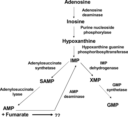
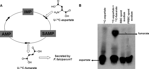
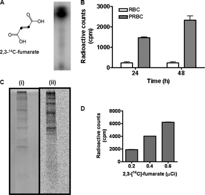
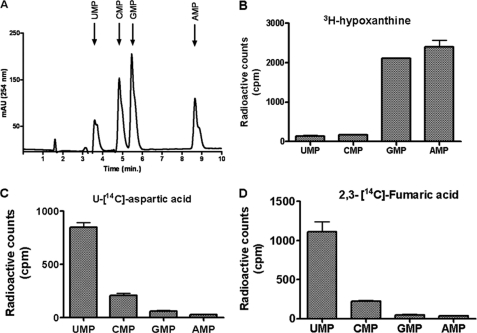
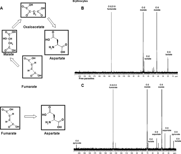
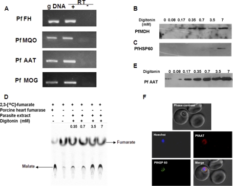
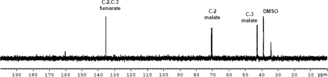
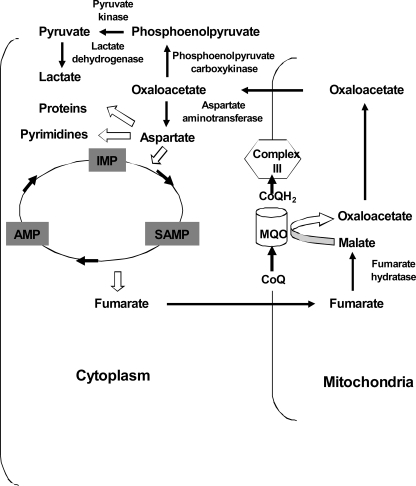
In aerobic respiration, the tricarboxylic acid cycle is pivotal to the complete oxidation of carbohydrates, proteins, and lipids to carbon dioxide and water. Plasmodium falciparum, the causative agent of human malaria, lacks a conventional tricarboxylic acid cycle and depends exclusively on glycolysis for ATP production. However, all of the constituent enzymes of the tricarboxylic acid cycle are annotated in the genome of P. falciparum, which implies that the pathway might have important, yet unidentified biosynthetic functions. Here we show that fumarate, a side product of the purine salvage pathway and a metabolic intermediate of the tricarboxylic acid cycle, is not a metabolic waste but is converted to aspartate through malate and oxaloacetate. P. falciparum-infected erythrocytes and free parasites incorporated [2,3-(14)C]fumarate into the nucleic acid and protein fractions. (13)C NMR of parasites incubated with [2,3-(13)C]fumarate showed the formation of malate, pyruvate, lactate, and aspartate but not citrate or succinate. Further, treatment of free parasites with atovaquone inhibited the conversion of fumarate to aspartate, thereby indicating this pathway as an electron transport chain-dependent process. This study, therefore, provides a biosynthetic function for fumarate hydratase, malate quinone oxidoreductase, and aspartate aminotransferase of P. falciparum.


Figures

FIGURE 1.
FIGURE 1.
Purine salvage pathway in the intraerythrocytic P. falciparum. Adenosine and hypoxanthine salvaged from the host are converted to IMP, which then branches out to form AMP and GMP. Excess AMP is deaminated back to IMP to constitute purine nucleotide cycle. A molecule of fumarate is also generated for each molecule of AMP. The arrow and the question mark have been put in to emphasize that the reutilization pathway of fumarate has not been elucidated.
FIGURE 2.
FIGURE 2.
Fumarate is not secreted as a metabolic waste by P. falciparum. A, the carbon skeleton of [U-14C]aspartate (in which all of the carbon atoms are enriched in 14C (shown as bold circles)) is converted to [U-14C]fumarate in the parasite by the concerted actions of adenylosuccinate synthetase and adenylosuccinate lyase. Arrows indicate the direction of the reaction. B, radio-TLC analysis of the spent medium of erythrocytes (2% hematocrit) and parasitized erythrocytes (2% hematocrit and 8% parasitemia) incubated with 1 μCi of [U-14C]aspartate for 24 h. [U-14C]Aspartate and a mixture of [U-14C]aspartate and [2,3-14C]fumarate were used as TLC markers. A solvent mixture of n-butanol, acetic acid, and water in the ratio of 4:1:1 was used as the mobile phase for silica-TLC. The TLC plate was dried and developed using a Fuji FLA-5000 phosphorimaging device.
FIGURE 3.
FIGURE 3.
[2,3-14C]fumarate incorporation into nucleic acids and proteins by parasitized erythrocytes and free parasites. A, structure and radio-TLC analysis of [2,3-14C]fumarate (10 mCi mmol−1) in which the second and third carbon atoms are enriched in 14C. B, [2,3-14C]fumarate incorporation by parasitized erythrocytes after incubation with the radioactive label for 24 and 48 h. 1 μCi of [2,3-14C]fumarate was added to PRBCs and RBCs, and cells were harvested and counted for radioactivity as described under “Experimental Procedures.” C, parasitized erythrocytes incubated with 1 μCi of [2,3-14C]fumarate for 24 h were treated with 0.15% saponin solution to release free parasites from erythrocytes. The parasite pellet was washed, processed, and subjected to SDS-PAGE, which was stained by Coomassie Brilliant Blue (i) and developed using a Fuji FLA-5000 phosphorimaging device (ii). D, radioactive incorporation in the nucleic acid fractions of saponin released erythrocyte-free parasites incubated with varying amounts of [2,3-14C]fumarate.
FIGURE 4.
FIGURE 4.
Purine and pyrimidine nucleotide labeling in free parasites by [8-3H]hypoxanthine, [U-14C]aspartate, and [2,3-14C]fumarate. A, separation of pyrimidine (UMP and CMP) and purine (GMP and AMP) mononucleotides on a C18 reversed phase HPLC. A mixture of nucleotide monophosphates was injected into the column and eluted with a linear gradient of solution B. B–D, soluble metabolites were extracted from free parasites that were incubated with 1 μCi of [3H]hypoxanthine (B), 1 μCi of [U-14C]aspartate (C), and 1 μCi of [2,3-14C]fumarate (D) and spiked with the nucleotide standards prior to HPLC. Peaks corresponding to the pyrimidine and purine nucleotide fractions were collected separately and concentrated, and incorporated radioactivity was measured as cpm using a Wallac 1409 liquid scintillation counter.
FIGURE 5.
FIGURE 5.
Metabolic intermediates in fumarate metabolism. A, pathways for fumarate metabolism. Fumarate can be converted to aspartate through the metabolic intermediates malate and oxaloacetate, or fumarate can be directly acted upon by aspartase to generate aspartate. B and C, RBCs (B) and PRBCs (C) incubated with 10 mm [2,3-13C]fumarate in 1× PBS at 37 °C for 4 h. Cell suspension was lysed and processed as described under “Experimental Procedures.” D2O was added to a final concentration of 10% as an internal lock to the cell lysate, and NMR spectra were recorded.
FIGURE 6.
FIGURE 6.
Expression and subcellular localization of enzymes involved in fumarate to aspartate conversion pathway. A, RT-PCR amplification of fragments of PfFH, PfMQO, PfAAT, and malate oxoglutarate (PfMOG) translocator. gDNA indicates reactions in which genomic DNA was used as a template for PCR. + and − RT indicate reactions with and without reverse transcriptase, respectively. −RT was included as a control to rule out genomic DNA contamination. B and C, Western blotting of the different subcellular fractions of P. falciparum parasites obtained with increasing concentrations of digitonin with anti-PfMDH and anti-PfHSP60 antisera. D, radio-TLC analysis for fumarate hydratase activity in different parasite cellular fractions obtained by digitonin treatment. Activity was assessed by the formation of [14C]malate from [2,3-14C]fumarate. The mobile phase consisted of a 4:1:1 mixture of n-butanol, acetic acid, and water. The silica-TLC plate was dried and developed using a Fuji FLA-5000 phosphorimaging device. E, Western blotting of subcellular fractions of P. falciparum by anti-PfAAT antisera. Low levels of PfAAT seen in the absence of digitonin are due to background lysis of cells. F, indirect immunofluorescence analysis of parasitized erythrocytes with anti-PfAAT and anti-PfHSP60 antisera. Anti-mouse and anti-rabbit IgG antibodies conjugated with Alexa Fluor dyes were used as secondary antibodies. Hoechst was used as the nuclear stain.
FIGURE 7.
FIGURE 7.
Atovaquone treatment inhibits the 13C enrichment of aspartate in parasites incubated with [2,3-13C]fumarate. Free parasites treated with 1 μm atovaquone for 2 h were then pulsed with 10 mm [2,3-13C]fumarate for 2 h. Following incubation, the parasites were processed as described under “Experimental Procedures.” DMSO, dimethyl sulfoxide.
FIGURE 8.
FIGURE 8.
Model for fumarate metabolism in P. falciparum. Cytosolic fumarate generated from the purine salvage pathway is metabolized to malate by mitochondrial fumarate hydratase, which then is converted to oxaloacetate by malate quinone oxidoreductase. In the latter step, CoQ is reduced to CoQH2, which is subsequently oxidized back to CoQ by complex III. Finally, oxaloacetate is converted to aspartate by a cytosolic aspartate aminotransferase.

References

    1. Bryant C., Voller A., Smith M. J. (1964) Am. J. Trop. Med. Hyg. 13, 515–519 - PubMed
    1. Kirk K., Horner H. A., Kirk J. (1996) Mol. Biochem. Parasitol. 82, 195–205 - PubMed
    1. Foth B. J., Stimmler L. M., Handman E., Crabb B. S., Hodder A. N., McFadden G. I. (2005) Mol. Microbiol. 55, 39–53 - PubMed
    1. Chen J., Jones M. (1976) Arch. Biochem. Biophys. 176, 82–90 - PubMed
    1. Wrenger C., Muller S. (2003) Eur. J. Biochem. 270, 1775–1783 - PubMed

Publication types

MeSH terms

LinkOut - more resources