. 2011 Mar;85(6):2953-63.
doi: 10.1128/JVI.02020-10. Epub 2011 Jan 5.

Subcellular localization and rearrangement of endoplasmic reticulum by Brome mosaic virus capsid protein

Affiliations

Subcellular localization and rearrangement of endoplasmic reticulum by Brome mosaic virus capsid protein

Devinka Bamunusinghe et al. J Virol. 2011 Mar.

Abstract

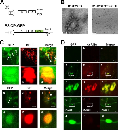
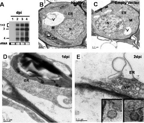
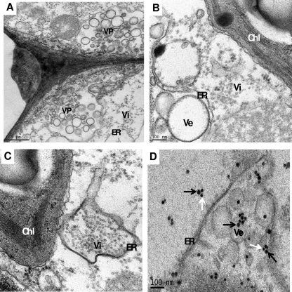
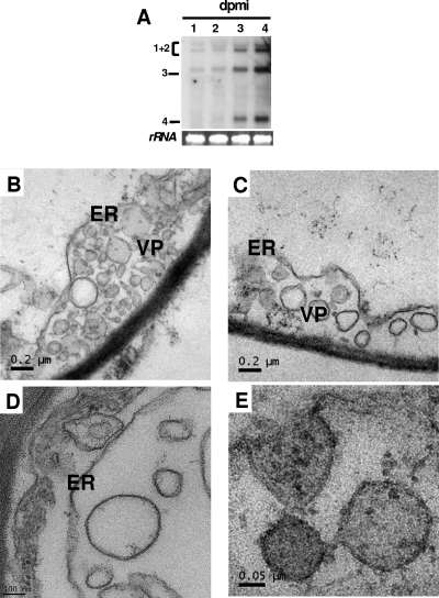
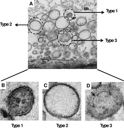
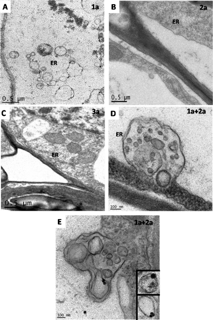
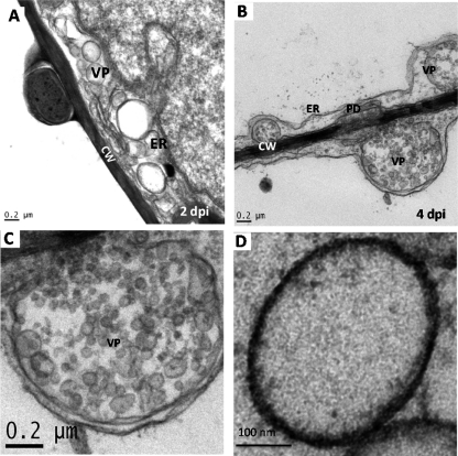
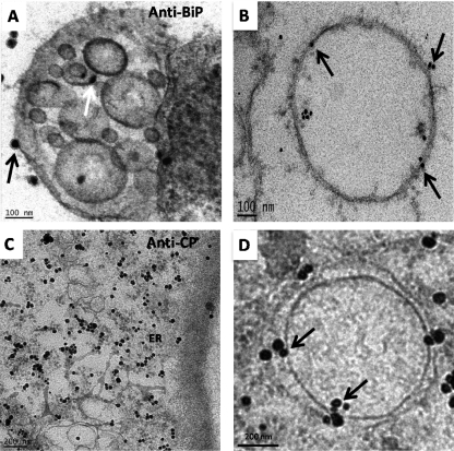
Genome packaging in the plant-infecting Brome mosaic virus (BMV), a member of the alphavirus-like superfamily, as well as in other positive-strand RNA viruses pathogenic to humans (e.g., poliovirus) and animals (e.g., Flock House virus), is functionally coupled to replication. Although the subcellular localization site of BMV replication has been identified, that of the capsid protein (CP) has remained elusive. In this study, the application of immunofluorescence confocal microscopy to Nicotiana benthamiana leaves expressing replication-derived BMV CP as a green fluorescent protein (GFP) fusion, in conjunction with antibodies to the CP and double-stranded RNA, a presumed marker of RNA replication, revealed that the subcellular localization sites of replication and CP overlap. Our temporal analysis by transmission electron microscopy of ultrastructural modifications induced in BMV-infected N. benthamiana leaves revealed a reticulovesicular network of modified endoplasmic reticulum (ER) incorporating large assemblies of vesicles derived from ER accumulated in the cytoplasm during BMV infection. Additionally, for the first time, we have found by ectopic expression experiments that BMV CP itself has the intrinsic property of modifying ER to induce vesicles similar to those present in BMV infections. The significance of CP-induced vesicles in relation to CP-organized viral functions that are linked to replication-coupled packaging is discussed.


Figures

FIG. 1.
FIG. 1.
Subcellular localization of BMV CP and replication sites. (A) Schematic representation of constructs used for in vitro transcription of wild-type BMV RNA3 (B3) and its chimera, B3/CP-GFP, designed to express GFP as a CP fusion protein. White boxes represent movement protein (3a) and CP ORFs. The position of the BamHI site used to linearize the plasmids for in vitro transcription using T7 polymerase is indicated. (B) TEM images of virions purified from N. benthamiana plants mechanically inoculated with the indicated inocula. (C) Subcellular localization of BMV CP. N. benthamiana leaves were mechanically inoculated with B1+B2+B3/CP-GFP inoculum and processed for IFCM. Antisera to ER-resident proteins KDEL and BiP were used to label ER followed by treatment with Alexa Fluor 633-conjugated secondary antibodies (emits red fluorescence). (Ca and Cd) Infected leaf cells showing aggregates of CP-GFP fusion as punctate bodies (indicated by arrows); (Cb and Ce) red fluorescence indicates ER; (Cc and Cf) yellow fluorescence in merged images represents where CP and ER colocalize; absence of yellow signal, representing noncolocalized areas, is indicated by arrowheads. Cellular regions boxed in panels a to c and d to f are shown enlarged in panels 1 to 3 and 4 to 6, respectively. (D) Colocalization of BMV CP and replication sites. Mock (Da to Dc)- or B1+B2+B3/CP-GFP (Dd to Df)-infected N. benthamiana leaves were probed for the presence of dsRNA using monoclonal antibody J2 and then treated with Alexa Fluor 633-conjugated secondary antibody. Green and red fluorescence in panels d and e, respectively, represent localization of the CP-GFP fusion protein and replication sites. Yellow fluorescence in panel f represents where CP and dsRNA colocalize. The cellular region boxed in panels d to f is shown enlarged in panels 1 to 3, respectively. (Dg and Dh) Infected leaves were treated with RNase III and probed for dsRNA. The cellular region boxed in panels g to i is shown enlarged in panels 4 to 6, respectively. Bars, 5 μm.
FIG. 2.
FIG. 2.
BMV replication correlates with vesicle induction in N. benthamiana leaves. (A) Temporal pattern of BMV RNA accumulation in N. benthamiana leaves infiltrated with agrotransformants of all three wt BMV RNAs. Northern blot showing the accumulation of BMV progeny RNA in leaves at 1 to 4 days postinfiltration (dpi). Five micrograms of total RNA per sample was subjected to fractionation on denaturing formaldehyde-agarose gels, transferred to a nylon membrane, and hybridized with a 32P-labeled riboprobe complementary to a sequence encompassing the conserved 3′ tRNA-like structure that detected all four BMV RNAs. The positions of the four BMV RNAs are indicated on the left. (B to E) BMV infection induces ultrastructural changes to the ER membrane. N. benthamiana leaves at various dpi were fixed, sectioned, and stained with uranyl acetate and analyzed by TEM. TEM images of a cell from healthy (B) and empty vector-infiltrated (C) leaves showing distribution of unmodified nucleus (N), endoplasmic reticulum (ER), vacuole (V), chloroplast (Chl), and mitochondria (M). (D) At 1 dpi, no change to any cellular organelle was observed. (E) A TEM image showing the formation of a vesicle packet (VP) containing a collection of virus-induced vesicles surrounded by ER at 2 dpi. (Insets) A magnified view of a vesicle as observed in many sections, showing a neck (dotted circle) connecting to ER. Bars, 0.5 μm.
FIG. 3.
FIG. 3.
Ultrastructure of distinct membrane alterations induced by BMV in N. benthamiana leaves at 4 dpi. A collection of TEM images of BMV-infected leaves fixed at 4 dpi are shown in panels A to C. (A) An overview of two neighboring cells infected by BMV showing a collection of virus-induced vesicles in the lumen of the ER to form a vesicle packet (VP). Assembled virions (Vi) are found scattered in the vicinity of VP. (B) An example of distinct cytopathological change induced by BMV infection showing accumulation of vesicles (Ve); (C) a higher-ordered ER structure accumulating assembled virions; (D) immunogold localization of viral replicase proteins 1a and 2a to vesicles in BMV-infected cells. Following fixation of leaf tissue, thin sections were labeled with antibodies specific to replicase proteins 1a and 2a using the SECSI technique (see Materials and Methods). Black and white arrows, respectively, represent 1a and 2a. Bars, 0.5 μm (A) and 100 nm (B to D).
FIG. 4.
FIG. 4.
Ultrastructure of membrane alterations induced by wt BMV infection in mechanically inoculated N. benthamiana leaves. (A) Northern blot showing the temporal pattern of BMV RNA accumulation at 1 to 4 days postmechanical inoculation (dpmi). (B to E) Representative TEM images showing the accumulation of vesicles in leaves at 3 dpmi. Bars, 0.2 μm (B and C), 100 nm (D), and 0.05 μm (E).
FIG. 5.
FIG. 5.
Types of vesicles associated with wt BMV infection. (A) An overview of vesicles accumulated in N. benthamiana leaves following agroinfiltration with wt BMV. Representative examples of three vesicle types, classified as type 1, 2, and 3, are indicated. Higher-magnified views of each vesicle type are shown in panels B to D. Note in panel D, the outer wall membrane is not continuous. An arrow indicates the internal structure with a single outer membrane. Bars, 200 nm.
FIG. 6.
FIG. 6.
Ectopic expression of BMV gene products. TEM images of leaf sections following ectopic expression of BMV replicase genes 1a (A) and 2a (B) and movement protein 3a (C) are shown. No change to any cellular organelle was observed in leaves infiltrated with either 2a (B) or 3a (C). (D) Accumulation of vesicles and formation of VP following coinfiltration with 1a and 2a. (E) Representative TEM image showing immunogold localization of dsRNA in vesicles induced by 1a plus 2a. (Inset) dsRNA localization on vesicles induced by 1a plus 2a from different tissue sections. Bars, 0.5 μm (A to C) and 100 nm (D and E).
FIG. 7.
FIG. 7.
Ectopic expression of BMV CP results in vesicle induction. TEM images showing induction of a collection of vesicles in N. benthamiana leaves ectopically expressing BMV CP at 2 dpi (A) and 4 dpi (B). A magnified view of a VP containing a collection of vesicles induced by CP in ER lumen is shown in panel C. A magnified view of a CP-induced vesicle is shown in panel D. Bars, 0.2 μm (A to C) and 100 nm (D).
FIG. 8.
FIG. 8.
Origin of CP-induced vesicles. (A) Leaf tissue ectopically expressing BMV CP was fixed and labeled with antibodies specific to BiP, an ER resident protein. Localization of BiP on the ER membrane surrounding CP-induced VP (black arrow) and on one of the vesicles (white arrow) is indicated. (B) An image showing a CP-induced vesicle labeled with BiP antibody (indicated by arrows). (C) Immunogold localization of anti-BMV CP in leaf tissue ectopically expressing BMV CP. (D) Magnified view of a CP-induced vesicle labeled with anti-BMV CP antibody (indicated by arrows) is shown. Bars, 100 nm (A and B) and 200 nm (C and D).

References

    1. Ahlquist, P. 2006. Parallels among positive-strand RNA viruses, reverse-transcribing viruses and double-stranded RNA viruses. Nat. Rev. Microbiol. 4:371-382. - PMC - PubMed
    1. Annamalai, P., and A. L. Rao. 2006. Delivery and expression of functional viral RNA genomes in planta by agroinfiltration, unit 16B, p. 2.1-2.15. In T. Downey (ed.), Current protocols in microbiology. John Wiley & Sons Inc., Mississauga, Ontario, Canada. - PubMed
    1. Annamalai, P., and A. L. Rao. 2006. Packaging of brome mosaic virus subgenomic RNA is functionally coupled to replication-dependent transcription and translation of coat protein. J. Virol. 80:10096-10108. - PMC - PubMed
    1. Annamalai, P., and A. L. Rao. 2005. Replication-independent expression of genome components and capsid protein of brome mosaic virus in planta: a functional role for viral replicase in RNA packaging. Virology 338:96-111. - PubMed
    1. Annamalai, P., F. Rofail, D. A. Demason, and A. L. Rao. 2008. Replication-coupled packaging mechanism in positive-strand RNA viruses: synchronized coexpression of functional multigenome RNA components of an animal and a plant virus in Nicotiana benthamiana cells by agroinfiltration. J. Virol. 82:1484-1495. - PMC - PubMed

Publication types

MeSH terms

Substances

LinkOut - more resources