. 2011 Jan 5;31(1):157-63.
doi: 10.1523/JNEUROSCI.1833-10.2011.

Novel regulation of parkin function through c-Abl-mediated tyrosine phosphorylation: implications for Parkinson's disease

Affiliations

Novel regulation of parkin function through c-Abl-mediated tyrosine phosphorylation: implications for Parkinson's disease

Syed Z Imam et al. J Neurosci. .

Abstract

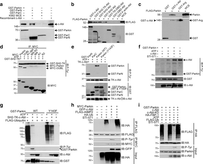
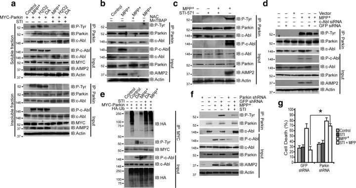
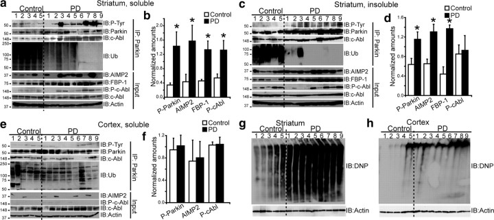
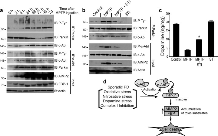
Mutations in parkin, an E3 ubiquitin ligase, are the most common cause of autosomal-recessive Parkinson's disease (PD). Here, we show that the stress-signaling non-receptor tyrosine kinase c-Abl links parkin to sporadic forms of PD via tyrosine phosphorylation. Under oxidative and dopaminergic stress, c-Abl was activated in cultured neuronal cells and in striatum of adult C57BL/6 mice. Activated c-Abl was found in the striatum of PD patients. Concomitantly, parkin was tyrosine-phosphorylated, causing loss of its ubiquitin ligase and cytoprotective activities, and the accumulation of parkin substrates, AIMP2 (aminoacyl tRNA synthetase complex-interacting multifunctional protein 2) (p38/JTV-1) and FBP-1.STI-571, a selective c-Abl inhibitor, prevented tyrosine phosphorylation of parkin and restored its E3 ligase activity and cytoprotective function both in vitro and in vivo. Our results suggest that tyrosine phosphorylation of parkin by c-Abl is a major post-translational modification that leads to loss of parkin function and disease progression in sporadic PD. Moreover, inhibition of c-Abl offers new therapeutic opportunities for blocking PD progression.


Figures

Figure 1.
Figure 1.
a, GST pull-down assay using GST-Parkin and parkin-fragments, GST-ParN and GST-ParC (∼1 μg each) with K562 whole-cell extracts (WCE) (5 mg). b, GST pull-down assay using GST-c-Abl and c-Abl fragments (∼1 μg) as shown in c and WCE (5 mg) of SH-SY5Y cells transfected with FLAG-parkin. *Degradation products of GST-c-Abl. c, GST pull-down assay using GST-Arg and GST-c-Abl (∼1 μg) and WCE (5 mg) of SH-SY5Y cells transfected with FLAG-parkin. d, Immunoblots with anti-p-tyrosine (P-Tyr) and myc after an in vitro kinase assay using GST-SH2-TK (c-Abl) and myc immunoprecipitates from SH-SY5Y cells transfected with myc-tagged full-length and deletion mutants of parkin (2 μg). e, Immunoblots with antibodies to P-Tyr, GST, c-Abl after an in vitro kinase assay using TK-c-Abl and GST-ParN, GST-ParC or GST-Parkin, wild-type or Y143F parkin. *Degradation products of GST-constructs. f, Immunoblots with antibodies to P-Tyr, GST, c-Abl after an in vitro kinase assay using GST-Parkin with full-length human recombinant His-c-Abl (100 ng) with or without STI-571 (2.5 μm). g, Immunoblots of in vitro auto-ubiquitination assays using SH2-TK-Abl (∼200 ng) and GST-Parkin (wild-type or Y143F). *Nonspecific ubiquitination. h, Immunoblot analysis of immunoprecipitates with FLAG and myc antibodies of SH-SY5Y cell lysates transfected with FLAG-tagged AIMP2 (2 μg), myc-tagged WT parkin (1 μg), HA-tagged ubiquitin (1 μg) and GFP-tagged c-Abl-KA (1 μg), incubated with 10 μm STI-571 for 12 h. i, Immunoblots of an in vitro GST-parkin ubiquitination assay of immunoprecipitated HA-FBP-1 with c-Abl or c-Abl KD (∼200 ng) with or without STI-571 (2.5 μm). All experiments were repeated at least three times. Representative examples are presented.
Figure 2.
Figure 2.
a, Immunoblots of parkin immunoprecipitates of SH-SY5Y TX-100-soluble (top) and TX-100-insoluble (bottom) fractions of cell lysates transiently transfected with myc-parkin treated with MPP+ (100 μm), DA (100 μm) for 24 h or with H2O2 (250 μm in serum free medium) for 1 h. In lanes 5, 6, and 7, cells were pretreated with STI-571 (STI) at 10 μm for 6 h before exposure to toxins. b, Immunoblots of parkin immunoprecipitates of SH-SY5Y cell lysates transiently transfected with myc-parkin treated with MPP+ (100 μm) for 24 h. In lanes 3 and 4, cells were pretreated with the superoxide dismutase mimetic MnTBAP (100 μm) and antioxidant NAC (1 mm) for 24 h before exposure to MPP+. c, Immunoblots of parkin immunoprecipitates of mouse primary striatal neurons (95% glia-free) treated with 100 nm MPP+ for 24 h with or without STI-571 (2.5 μm for 6 h before MPP+ treatment). d, Immunoblots of parkin immunoprecipitates of SH-SY5Y cells transiently transfected with c-Abl siRNA or GFP siRNA 48 h before MPP+ (100 μm) treatment. e, Immunoblots of myc immunoprecipitates of SH-SY5Y cell lysates transiently transfected with myc-parkin and HA-ubiquitin and treated with MPP+ or DA (100 μm) for 24 h. In lanes 5 and 6, cells were pretreated with STI-571 at 10 μm for 6 h before exposure to MPP+ or DA. f, Immunoblots of parkin immunoprecipitates of SH-SY5Y cells, infected with either lenti-shRNA to parkin or lenti-shRNA to GFP, treated with 100 μm MPP+. Some samples were incubated with 10 μm STI-571 for 6 h before MPP+ treatment. g, Cell death plotted as percentage of PI-positive cells among GFP-positive cells of SH-SY5Y cells infected with lenti-shRNA-parkin or lenti-shRNA-GFP alone 48 h before treatment with MPP+ (100 μm). Some samples were incubated with 10 μm STI-571 for 6 h before MPP+ treatment. *p < 0.05. Differences among means were analyzed using one-way ANOVA with the different treatments as the independent factor followed by Newman–Keuls post hoc analysis. All experiments were repeated at least three times. Representative examples are presented.
Figure 3.
Figure 3.
a, c, e, Immunoblots of parkin immunoprecipitates (2 mg) prepared from lysates of TX-100-soluble striatum (a), TX-100-insoluble striatum (c), and TX-100-soluble cortex lysates (e). Brain tissue lysates (100 μg) were immunoblotted with antibodies to parkin, AIMP2, FBP-1, ubiquitin (Ub), P-tyr, P-c-Abl (p-Y245), c-Abl, and actin. b, d, f, Ratios of phospho-parkin/IP parkin and AIMP2/actin from lysates of TX-100-soluble striatum (b), TX-100-insoluble striatum (d), and TX-100-soluble cortex for control versus PD (f) (*p < 0.05, Student's t test). g, h, OxyBlot analysis of proteins from postmortem striatum (g), and cortex of PD patients and age-matched controls (h). All experiments were repeated at least three times. Representative examples are presented.
Figure 4.
Figure 4.
a, Immunoblots of c-Abl and parkin IP samples (2 mg) from striatum tissue lysates of saline- or MPTP-treated mice (4 doses of 20 mg/kg MPTP, i.p., at 2 h intervals at indicated time points). b, Immunoblots of parkin immunoprecipitates of striatum lysates (100 μg) 7 d after MPTP treatment with or without STI-571(STI) (10 mg/kg, i.p.), (daily injection for 7 d before and 7 d after MPTP injection) compared with saline- and STI-injected control mice. c, STI-571 prevents MPTP-induced depletion of striatal dopamine in mice, as assessed by HPLC/electrochemical detection. Each value is mean ± SEM derived from 10 animals/group. Statistical significance was assessed using one-way ANOVA and Student–Newman–Keuls multiple-comparisons test. *p < 0.05 for difference from MPTP group. d, Schematic representation of the pathway by which parkin tyrosine phosphorylation can lead to cell death and PD. Oxidative or dopamine stress from an external source or generated during sporadic PD activates c-Abl. Activated c-Abl tyrosine phosphorylates parkin resulting in loss of ubiquitin ligase activity, leading to accumulation of toxic parkin substrates and neuronal death.

References

    1. Braak H, Del Tredici K, Rüb U, de Vos RA, Jansen Steur EN, Braak E. Staging of brain pathology related to sporadic Parkinson's disease. Neurobiol Aging. 2003;24:197–211. - PubMed
    1. Breedveld P, Pluim D, Cipriani G, Wielinga P, van Tellingen O, Schinkel AH, Schellens JH. The effect of Bcrp1 (Abcg2) on the in vivo pharmacokinetics and brain penetration of imatinib mesylate (Gleevec): implications for the use of breast cancer resistance protein and P-glycoprotein inhibitors to enable the brain penetration of imatinib in patients. Cancer Res. 2005;65:2577–2582. - PubMed
    1. Chung KK, Zhang Y, Lim KL, Tanaka Y, Huang H, Gao J, Ross CA, Dawson VL, Dawson TM. Parkin ubiquitinates the alpha-synuclein-interacting protein, synphilin-1: implications for Lewy-body formation in Parkinson disease. Nat Med. 2001;7:1144–1150. - PubMed
    1. Chung KK, Thomas B, Li X, Pletnikova O, Troncoso JC, Marsh L, Dawson VL, Dawson TM. S-nitrosylation of parkin regulates ubiquitination and compromises parkin's protective function. Science. 2004;304:1328–1331. - PubMed
    1. Corti O, Hampe C, Koutnikova H, Darios F, Jacquier S, Prigent A, Robinson JC, Pradier L, Ruberg M, Mirande M, Hirsch E, Rooney T, Fournier A, Brice A. The p38 subunit of the aminoacyl-tRNA synthetase complex is a Parkin substrate: linking protein biosynthesis and neurodegeneration. Hum Mol Genet. 2003;12:1427–1437. - PubMed

Publication types

MeSH terms

Substances