

Central amygdala activity during fear conditioning
- PMID: 21209214
- PMCID: PMC3080118
- DOI: 10.1523/JNEUROSCI.4985-10.2011
Central amygdala activity during fear conditioning
Abstract
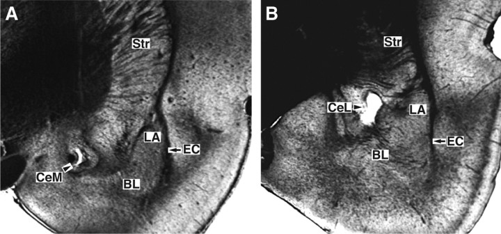
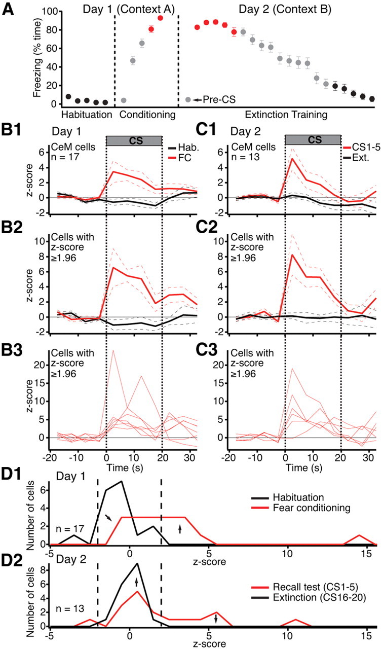
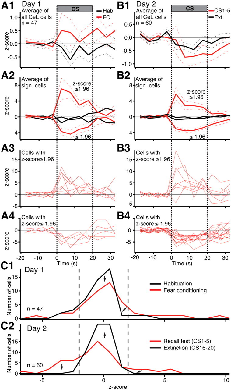
The central amygdala (Ce), particularly its medial sector (CeM), is the main output station of the amygdala for conditioned fear responses. However, there is uncertainty regarding the nature of CeM control over conditioned fear. The present study aimed to clarify this question using unit recordings in rats. Fear conditioning caused most CeM neurons to increase their conditioned stimulus (CS) responsiveness. The next day, CeM cells responded similarly during the recall test, but these responses disappeared as extinction of conditioned fear progressed. In contrast, the CS elicited no significant average change in central lateral (CeL) firing rates during fear conditioning and a small but significant reduction during the recall test. Yet, cell-by-cell analyses disclosed large but heterogeneous CS-evoked responses in CeL. By the end of fear conditioning, roughly equal proportions of CeL cells exhibited excitatory (CeL(+)) or inhibitory (CeL(-)) CS-evoked responses (∼10%). The next day, the proportion of CeL(-) cells tripled with no change in the incidence of CeL(+) cells, suggesting that conditioning leads to overnight synaptic plasticity in an inhibitory input to CeL(-) cells. As in CeM, extinction training caused the disappearance of CS-evoked activity in CeL. Overall, these findings suggest that conditioned freezing depends on increased CeM responses to the CS. The large increase in the incidence of CeL(-) but not CeL(+) cells from conditioning to recall leads us to propose a model of fear conditioning involving the potentiation of an extrinsic inhibitory input (from the amygdala or elsewhere) to CeL, ultimately leading to disinhibition of CeM neurons.
Figures
References
-
- Amano T, Duvarci S, Pare D. The basal nuclei of the amygdala are involved in conditioned fear extinction but not expression. Soc Neurosci Abstr. 2010;36:808–24.
-
- Cassell MD, Gray TS, Kiss JZ. Neuronal architecture in the rat central nucleus of the amygdala: a cytological, hodological, and immunocytochemical study. J Comp Neurol. 1986;246:478–499. - PubMed
-
- Ciocchi S, Herry C, Grenier F, Wolff SB, Letzkus JJ, Vlachos I, Ehrlich I, Sprengel R, Deisseroth K, Stadler MB, Müller C, Lüthi A. Encoding of conditioned fear in central amygdala inhibitory circuits. Nature. 2010;468:277–282. - PubMed
-
- Davis M. The role of the amygdala in conditioned and unconditioned fear and anxiety. In: Aggleton JP, editor. The amygdala. Oxford: Oxford UP; 2000. pp. 213–287.
Publication types
MeSH terms
Grants and funding
LinkOut - more resources
Full Text Sources