Comparative Study
. 2011 Feb 1;186(3):1575-88.
doi: 10.4049/jimmunol.1002990. Epub 2011 Jan 5.

Although divergent in residues of the peptide binding site, conserved chimpanzee Patr-AL and polymorphic human HLA-A*02 have overlapping peptide-binding repertoires

Affiliations
Comparative Study

Although divergent in residues of the peptide binding site, conserved chimpanzee Patr-AL and polymorphic human HLA-A*02 have overlapping peptide-binding repertoires

Michael Gleimer et al. J Immunol. .

Abstract

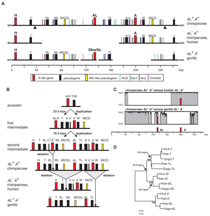
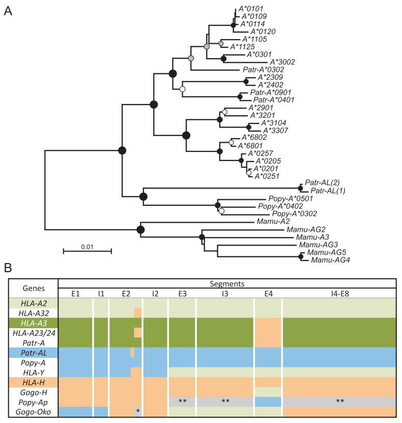
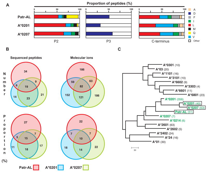
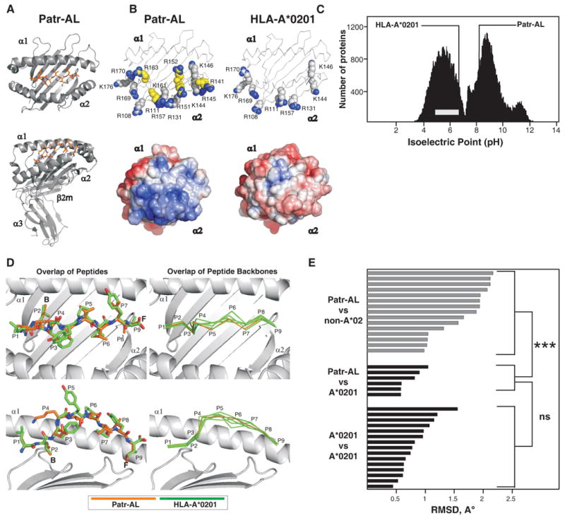
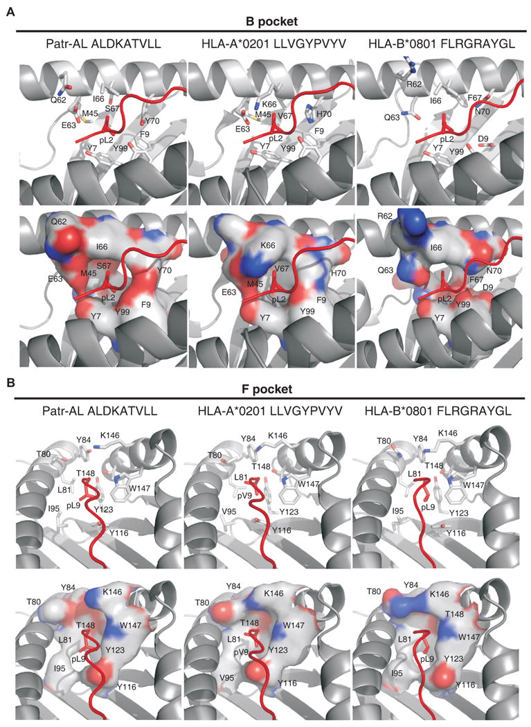
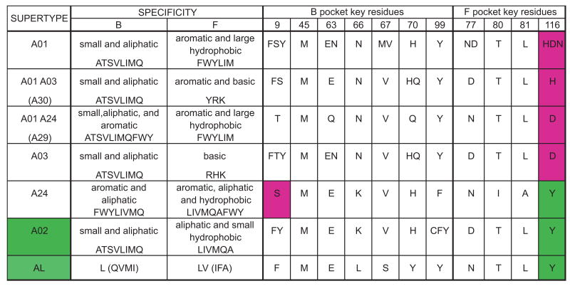
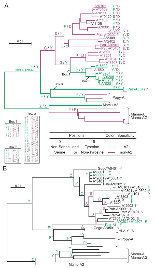
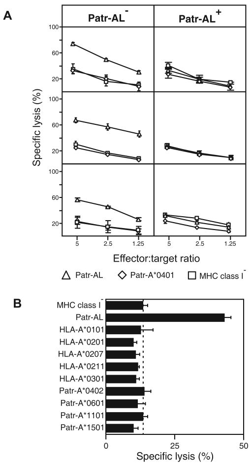
Patr-AL is an expressed, non-polymorphic MHC class I gene carried by ∼50% of chimpanzee MHC haplotypes. Comparing Patr-AL(+) and Patr-AL(-) haplotypes showed Patr-AL defines a unique 125-kb genomic block flanked by blocks containing classical Patr-A and pseudogene Patr-H. Orthologous to Patr-AL are polymorphic orangutan Popy-A and the 5' part of human pseudogene HLA-Y, carried by ∼10% of HLA haplotypes. Thus, the AL gene alternatively evolved in these closely related species to become classical, nonclassical, and nonfunctional. Although differing by 30 aa substitutions in the peptide-binding α(1) and α(2) domains, Patr-AL and HLA-A*0201 bind overlapping repertoires of peptides; the overlap being comparable with that between the A*0201 and A*0207 subtypes differing by one substitution. Patr-AL thus has the A02 supertypic peptide-binding specificity. Patr-AL and HLA-A*0201 have similar three-dimensional structures, binding peptides in similar conformation. Although comparable in size and shape, the B and F specificity pockets of Patr-AL and HLA-A*0201 differ in both their constituent residues and contacts with peptide anchors. Uniquely shared by Patr-AL, HLA-A*0201, and other members of the A02 supertype are the absence of serine at position 9 in the B pocket and the presence of tyrosine at position 116 in the F pocket. Distinguishing Patr-AL from HLA-A*02 is an unusually electropositive upper face on the α(2) helix. Stimulating PBMCs from Patr-AL(-) chimpanzees with B cells expressing Patr-AL produced potent alloreactive CD8 T cells with specificity for Patr-AL and no cross-reactivity toward other MHC class I molecules, including HLA-A*02. In contrast, PBMCs from Patr-AL(+) chimpanzees are tolerant of Patr-AL.


Figures

Figure 1
Figure 1. The MHC-AL+ haplotype arose through two MHC class I block duplications and one deletion, and gave rise to other hominid MHC haplotypes through deletion of either the MHC-A or MHC-AL blocks
(A) Compares the genomic organization of the chimpanzee AL+A+ haplotype to human (GenBank ID BA000025) and chimpanzee AL- A+ haplotypes (GenBank ID BA000041), and to the gorilla AL+A- haplotype (GenBank IDs CU104658, CU104664). Genes, pseudogenes, gene fragments, and informative repetitive elements are shown. (B) Model for the evolution through duplication and deletion of MHC haplotypes containing the H, AL, and A genes. U and A are hypothetical paralogs that have yet to be indentified in any hominoid species or on any sequenced MHC haplotype. (C) Shows the sequence similarity between the chimpanzee AL+A+ haplotype and both the human AL- A+ (upper panel) and gorilla AL+A- (lower panel) haplotypes. A sliding window of 100 bp was analyzed. Positions of the AL and A genes are indicated. (D) Shows a neighbor-joining phylogenetic tree of the genomic segments containing the pseudogenes related to HLA-T, HLA-W, and HLA-K that was used to estimate the time of the two key duplication events. Arrows show the nodes used for maximum likelihood time estimates of the duplications.
Figure 2
Figure 2. Patr-AL is orthologous to orangutan Popy-A and to 3′ segments of human HLA-Y and gorilla Gogo-Oko
(A) Phylogenetic reconstruction was performed using neighbor-joining, maximum-likelihood, and maximum-parsimony methods. Shown is the neighbour-joining tree, with midpoint rooting and black, grey and white circles indicating ≥95, 80 and 60% bootstrap support, respectively, with all three methods. For the analysis exon 4, encoding the α3 domain, and the non-coding regions in the 5′ and 3′ of the gene were excluded. (B) Schematic showing the phylogenetic relationships between the MHC-A, MHC-H and MHC-AL and related MHC class I gene sequences. Equivalent segments between or within species share the same colors. These conclusions are based on the results from domain-by-domain phylogenetic analyses, and recombination detection methods (see Methods). HLA-A2 and HLA-A3 refer to the two major lineages of HLA-A alleles. *, MHC-A/AL group. **, outgroup to both MHC-A and -AL. Mamu, Macaca mulatta; Popy, Pongo pygmaeus; Patr, Pan troglodytes; Gogo, Gorilla gorilla.
Figure 3
Figure 3. Extensive overlap between the repertoires of peptides bound by Patr-AL and HLA-A*02
(A) Distribution of amino acid residues at the three anchor positions of the bound peptide: position 2 (P2), position 3 (P3), and the C-terminus. Proportions were calculated from the amino-acid sequences of 126 binding peptides for Patr-AL, HLA-A*0201 and HLA-A*0207. (B) Overlap of the peptide repertoires bound by Patr-AL, HLA-A*0201 and HLA-A*0207. The peptides binding to each MHC class I are defined by the differently colored circles: Patr-AL, red; A*0201, blue; and A*0207, green. The four overlapping regions between the circles defines the peptides bound by all three MHC class I and by the three combinations of two of them. On the left under ‘Sequenced peptides’ is shown the analysis for the peptides for which the amino-acid sequences are known. On the right under ‘Molecular ions’ is shown a second, independent study in which peptides were defined by the weight of their molecular ions. (C) Neighbor-joining phylogenetic tree to compare the peptide-binding specificities of Patr-AL, A*0201, and A*0207 as defined in our analysis (green and boxed), with those previously defined for A*0201, A*0207, A*0214 (green) and sixteen other HLA-A allotypes obtained from the SYFPEITHI database (39). The number of peptides in each dataset is shown in parentheses following the allotype name.
Figure 4
Figure 4. Despite their distinctive molecular surfaces, Patr-AL and HLA-A*0201 bind peptides in similar conformation
(A) Ribbon diagrams of the crystallographic structure of the complex of peptide ALDKATVLL (colored red) bound to Patr-AL. The upper diagram is a top view showing the peptide bound by the α1and α2 domains. The lower diagram is a side view showing all four extracellular Patr-AL domains.(B) Compares the distribution of electropositive residues on the top face of Patr-AL (PDB ID pending) and HLA-A*0201(PDB ID 1HHK). The upper diagrams show the position of positively-charged residues; those unique to Patr-AL colored yellow. In the lower diagrams, Poisson-Boltzmann electrostatic potentials of Patr-AL and HLA-A*0201 are projected onto solvent-exposed surfaces and colored from red (-5.0) to blue (+5.0). (C) Compares the isoelectric point of Patr-AL to those of other cellular proteins. The bimodal black-shaded distribution represents all cellular proteins (88), the grey bar shows the range of isoelectric point for MHC class I other than Patr-AL. The isoelectric points of Patr-AL and HLA-A*02 are indicated by the lines. (D) Visual comparison of the conformation of peptide bound by Patr-AL and HLA-A*020. The left panels show least-squares superimposition of Patr-AL and HLA-A*0201 peptide-binding domains, which results in close alignment of peptide Cα backbones and similar side chain orientations. The positions of peptide residues and pockets B and F are indicated. The right panels show alignment of the Patr-AL bound peptide backbone with its counterparts in six structures of different peptides bound to HLA-A*0201 (PDB IDs 1HHK, 1QEW, 1B0G, 1HHI, 1HHG, 1HHJ). (E) Quantitative comparison of the conformation of peptide bound to Patr-AL and HLA class I. RMSDs of the Cα backbone for Patr-AL bound peptides fall within the range observed for HLA-A*0201 peptides, and are significantly lower than for peptides bound to non-A*02 HLA class I. structures. The details of the allotypes and peptides compared are in supplementary Fig. S5.
Figure 5
Figure 5. Patr-AL and HLA-A*0201 (1HHK) but not HLA-B*0801 (1M05) share binding site architecture despite non-conservative changes in specificity-determining pockets B and F
The architecture of (A) the B pocket and (B) the F pocket in the structures of Patr-AL, HLA-A*0201 (1HHK) and HLA-B*0801 (1M05) in complexes with peptide are compared. Peptide is shown in red. Top panels, stick representations of side chains forming the pockets are shown in gray; bottom panels, molecular surfaces of the pockets are shown. Residues and approximate positions of the B and F pockets are indicated.
Figure 6
Figure 6. The A02 supertype can be simply described by the residues at positions 9 and 116 of the peptide binding domain
Shown are the supertypes defined by Sidney et al (68), the characteristics of the residues accommodated by the B and F pockets and the amino acids found at the key residues of these pockets for all allotypes. All allotypes assigned to a supertype were aligned and the amino acids present at the key residue positions were determined; they are shown using the single letter code. Only A02 and A24 can accommodate aliphatic residues in the F pocket, a feature correlating with the presence of tyrosine at position 116 of the peptide-binding domain (highlighted green in the figure). They differ in the residues that can be accommodated in the B pocket, with A24 able to accommodate aromatic residues, a distinction that correlates with the presence of serine at position 9 of the peptide-binding domain (highlighted purple).
Figure 7
Figure 7. Evolution of the A2 peptide-binding specificity at the HLA-A gene
Combination of residues at positions 9 and 116 of the MHC-A and -AL peptide binding domain correlates with the presence or absence of the A2 peptide specificity (summarized in Fig. 6). Panel A shows the results of ancestral sequence reconstruction for both positions on the phylogenetic tree shown in Fig. 2. To complement this analysis, and extend the set of sequences that could be included in the analysis, we generated a phylogenetic tree on a smaller gene segment beginning 300bp upstream of the ATG start codon and ending in exon 2 (supplemental Fig. S6) and used this tree for ancestral reconstruction of position 9 (panel B). (A) The identities at positions 9 and 116 from the ancestral sequence reconstruction are shown for thirteen nodes. The branches of the tree and the names of the sequences are colored according to peptide specificity (green for A02 and purple for non-A02): for the branches, peptide specificity is based on the residues predicted at positions 9 and 116. For contemporary allotypes, the specificities were obtained from Sidney et al. (68) (underlining indicates ‘observed’ specificities). For each sequence the residues at positions 9 and 116 are shown to the right. The amino acid changes that occurred along three branches are given in boxes: positions colored in blue represent peptide-binding sites (89), residues colored in red along the HLA-A2 branches represent residues found in Patr-AL, while residues colored in red along the Patr-AL branch represent HLA-A*0201 residues. Underlined residues have p>0.95. #, specificity is based on Patr-A*0301. (B) Identity of position 9 from the ancestral sequence reconstruction is shown for all nodes. For each sequence the residue at position 9 is shown to the right. Serine is colored purple, all others are green. Names of MHC class I with A02 peptide specificity are green. Underlined residues have p>0.95. *, non-serine residue (p=1.0).
Figure 8
Figure 8. Patr-AL is an alloantigen that stimulates highly specific CD8 T cells
(A) Stimulating PBMC from Patr-AL- chimpanzees with Patr-AL expressing transfected 221 cells led to the generation of Patr-AL specific CD8+ cytotoxic T cell lines (CTL). As shown for CTL produced from PBMC of three individual Patr-AL- chimpanzees (left panels), these T cells killed 221 target cells expressing Patr-AL but not 221 cells expressing either Patr-A*0401 or untransfected 221 cells. In contrast, when PBMC from three Patr-AL+ chimpanzees were stimulated with 221 cells expressing Patr-AL, no CTL reactive with Patr-AL were produced (right panels). Patr-AL is thus seen to be an alloantigen for Patr-AL- individuals and to generate T cell tolerance in Patr-AL+ individuals. (B) Shown are the results of cytotoxicity assays performed with Patr-AL specific CTL from one individual chimpanzee and transfected 221 target cells expressing a range of human and chimpanzee MHC-A allotypes. The CTL are exquisitely specific for Patr-AL, exhibiting no significant cross-reactivity with any of the MHC-A variants tested. Assays shown were performed with an effector:target cell ratio of 2.5. Data in A and B are shown as mean ± SD for triplicates and represent at least two independent experiments.

References

    1. Peaper DR, Cresswell P. Regulation of MHC class I assembly and peptide binding. Annu Rev Cell Dev Biol. 2008;24:343–368. - PubMed
    1. Cheent K, Khakoo SI. Natural killer cells: integrating diversity with function. Immunology. 2009;126:449–457. - PMC - PubMed
    1. Jenkins MK, Chu HH, McLachlan JB, Moon JJ. On the composition of the preimmune repertoire of T cells specific for Peptide-major histocompatibility complex ligands. Annu Rev Immunol. 2010;28:275–294. - PubMed
    1. Adams EJ, Parham P. Species-specific evolution of MHC class I genes in the higher primates. Immunol Rev. 2001;183:41–64. - PubMed
    1. Shiina T, Ota M, Shimizu S, Katsuyama Y, Hashimoto N, Takasu M, Anzai T, Kulski JK, Kikkawa E, Naruse T, Kimura N, Yanagiya K, Watanabe A, Hosomichi K, Kohara S, Iwamoto C, Umehara Y, Meyer A, Wanner V, Sano K, Macquin C, Ikeo K, Tokunaga K, Gojobori T, Inoko H, Bahram S. Rapid evolution of major histocompatibility complex class I genes in primates generates new disease alleles in humans via hitchhiking diversity. Genetics. 2006;173:1555–1570. - PMC - PubMed

Publication types

MeSH terms