. 2010 Jan 26;3(1):127-35.
doi: 10.1007/s12307-010-0036-5.

Cancer-stromal interactions in scirrhous gastric carcinoma

Cancer-stromal interactions in scirrhous gastric carcinoma

Masakazu Yashiro et al. Cancer Microenviron. .

Abstract

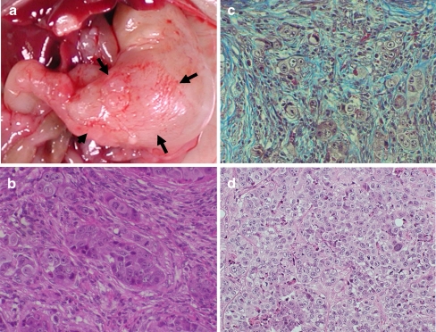
Fibroblasts play an important role in the progression, growth and spread of gastric cancers. Cancer-stroma interactions have been especially evident in the scirrhous type of gastric carcinoma. Fibroblasts are associated with the cancer progression at the primary and metastatic site. The proliferative and invasive ability of scirrhous gastric cancer cells are closely associated with the growth factors produced by organ-specific fibroblasts. Fibroblasts are therefore a key determinant in the malignant progression of gastric cancer and represent an important target for cancer therapies.

Keywords: Fibroblasts; Growth factor; Interaction; Microenvironment; Scirrhous gastric cancer; Stromal cells.


Figures

Fig. 1
Fig. 1
Scirrhous gastric cancer. a Macroscopic findings indicate that scirrhous gastric carcinoma diffusely infiltrates a broad region of the stomach without a prominent ulceration and elevation. b Microscopical views of scirrhous gastric carcinoma have the appearance of abundant fibrosis with the occasional presence of poorly differentiated cancer cells
Fig. 2
Fig. 2
Interaction between gastric fibroblasts and scirrhous gastric cancer cells. a The tumor size achieved by the co-inoculation of scirrhous gastric cancer cells and gastric fibroblasts was much larger than that achieved by the inoculation of cancer cells alone. b The xenografts produced by the co-inoculation shows infiltrative growth with extensive fibrosis, while the xenografted tumor of scirrhous gastric cancer cells alone shows medullary growth with poor fibrosis
Fig. 3
Fig. 3
Orthotopic implantation. a Macroscopic findings of the gastric tumor after orthotopic inoculation of gastric cancer cells showed scirrhous type carcinoma. b The orthotopic tumors showed extensive fibrosis with the presence of cancer cells which resemble human scirrhous gastric carcinoma. c Masson trichrome staining showed blue-colored multiple fibrosis in the orthotopic tumors. d In contrast, the histological findings of a subcutaneous tumor by scirrhous gastric carcinoma cells showed medullary growth with less fibrosis (e)
Fig. 4
Fig. 4
Cancer–stroma interaction of scirrhous gastric carcinoma at primary sites. At early stage, tumor cells need to invade into submucosa beyond the mucularis mucosae. MMP-2 production from fibroblasts and loss of cell–cell adhesion by down-regulation of E-cadherin and Desmoglein-2 might be important for the invasion of scirrhous cancer cells. When the tumor cells invade into the submucosa, a close relationship between cancer cells and orthotopic fibroblasts was evident in the development of gastric scirrhous cancer. Fibroblast growth factor 7 (FGF-7); a ligand of FGFR-2, secreted from gastric fibroblasts is likely to contribute in a paracrine manner to the remarkable cell proliferation seen in gastric cancer with FGF-R2 amplification. TGFβ or HGF produced from stromal fibroblasts stimulates the invasion ability of scirrhous gastric cancer cells. TGFβ from gastric cancer cells stimulates the proliferation of fibroblasts. FGF-7, TGFβ and HGF organ-specific fibroblasts and TGFβ from gastric cancer cells could mutually increase each other’s proliferation, thus resulting in the characteristic histology of scirrhous gastric carcinoma
Fig. 5
Fig. 5
Cancer–stroma interactions in scirrhous gastric carcinoma at peritoneal metastatic sites. Cancer cells free in the abdominal cavity initially adhere to hyaluronic acid on the mesothelial cells mediated by CD44 on the cancer cells. Thereafter, cancer cells adhere to submesothelial matrix of laminin, fibronectin, and collagen by α2β1- and α3β1-integrin expressed on cancer cells. Gastric cancer cells leaving the primary tumor are exposed to low oxygen levels in the peritoneal cavity. Hypoxia increases the expression of integrin by the cancer cells. Peritoneal fibroblasts are induced by cancer cells to morphologically change into mesothelial cells, and stimulate the migratory capability of cancer cells by TGF-β, HGF and MMPs. Peritoneal fibrosis induced by cancer cells prior to cancer metastases may create a congenial environmental “soil” for peritoneal metastases of scirrhous gastric carcinoma

References

    1. Kalluri R, Zeisberg M. Fibroblasts in cancer. Nat Rev Cancer. 2006;6(5):392–401. doi: 10.1038/nrc1877. - DOI - PubMed
    1. Japanese Gastric Cancer A Japanese classification of gastric carcinoma—2nd English Edition. Gastric Cancer. 1998;1(1):10–24. doi: 10.1007/PL00011681. - DOI - PubMed
    1. Ikeguchi M, Miyake T, Matsunaga T, et al. Recent results of therapy for scirrhous gastric cancer. Surg Today. 2009;39(4):290–294. doi: 10.1007/s00595-008-3860-1. - DOI - PubMed
    1. Otsuji E, Kuriu Y, Okamoto K, et al. Outcome of surgical treatment for patients with scirrhous carcinoma of the stomach. Am J Surg. 2004;188(3):327–332. doi: 10.1016/j.amjsurg.2004.06.010. - DOI - PubMed
    1. Tahara E. Genetic pathways of two types of gastric cancer. IARC Sci Publ. 2004;157:327–349. - PubMed

LinkOut - more resources