. 2010 Nov 30;5(11):e14154.
doi: 10.1371/journal.pone.0014154.

Reduced expression of the ROCK inhibitor Rnd3 is associated with increased invasiveness and metastatic potential in mesenchymal tumor cells

Affiliations

Reduced expression of the ROCK inhibitor Rnd3 is associated with increased invasiveness and metastatic potential in mesenchymal tumor cells

Cristina Belgiovine et al. PLoS One. .

Abstract

Background: Mesenchymal and amoeboid movements are two important mechanisms adopted by cancer cells to invade the surrounding environment. Mesenchymal movement depends on extracellular matrix protease activity, amoeboid movement on the RhoA-dependent kinase ROCK. Cancer cells can switch from one mechanism to the other in response to different stimuli, limiting the efficacy of antimetastatic therapies.

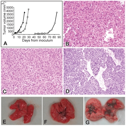
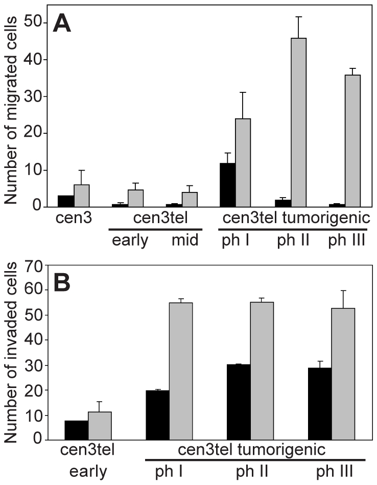
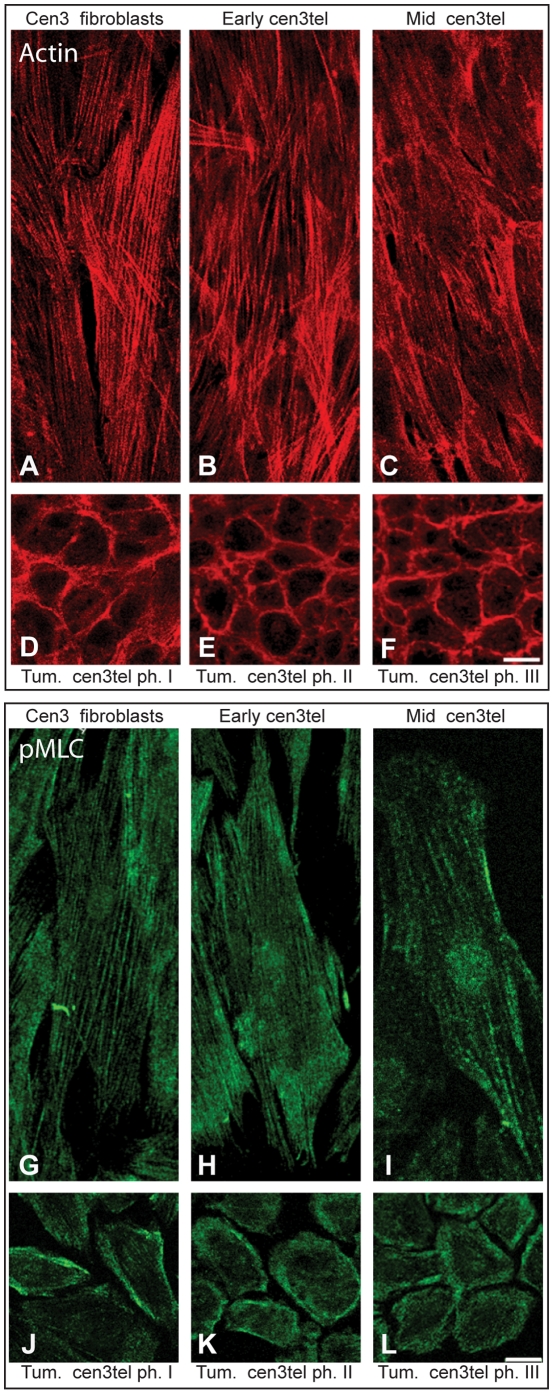
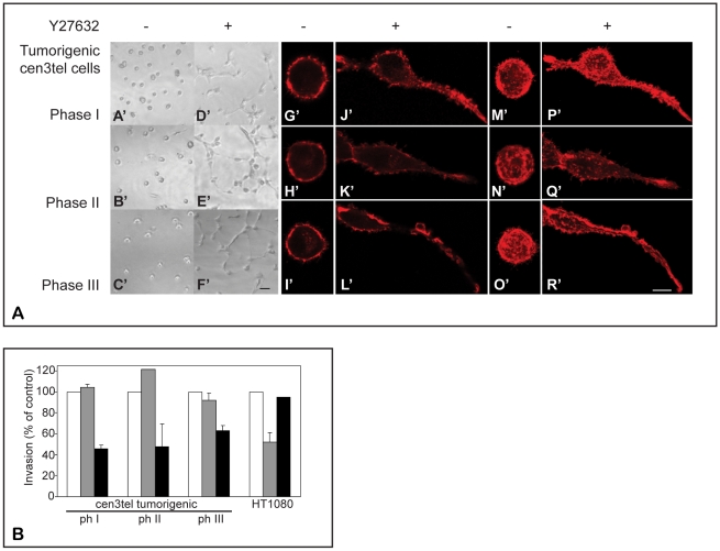
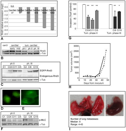
Methodology and principal findings: We investigated the acquisition and molecular regulation of the invasion capacity of neoplastically transformed human fibroblasts, which were able to induce sarcomas and metastases when injected into immunocompromised mice. We found that neoplastic transformation was associated with a change in cell morphology (from fibroblastic to polygonal), a reorganization of the actin cytoskeleton, a decrease in the expression of several matrix metalloproteases and increases in cell motility and invasiveness. In a three-dimensional environment, sarcomagenic cells showed a spherical morphology with cortical actin rings, suggesting a switch from mesenchymal to amoeboid movement. Accordingly, cell invasion decreased after treatment with the ROCK inhibitor Y27632, but not with the matrix protease inhibitor Ro 28-2653. The increased invasiveness of tumorigenic cells was associated with reduced expression of Rnd3 (also known as RhoE), a cellular inhibitor of ROCK. Indeed, ectopic Rnd3 expression reduced their invasive ability in vitro and their metastatic potential in vivo.

Conclusions: These results indicate that, during neoplastic transformation, cells of mesenchymal origin can switch from a mesenchymal mode of movement to an amoeboid one. In addition, they point to Rnd3 as a possible regulator of mesenchymal tumor cell invasion and to ROCK as a potential therapeutic target for sarcomas.


Conflict of interest statement

Competing Interests: The authors have declared that no competing interests exist.

Figures

Figure 1
Figure 1. Tumor induction by cen3tel cells.
A) Growth curves of tumors induced by cen3tel cells at PD 165 (empty circles), PD 616 (empty squares) and PD 902 (black triangles). B–D) Histological analysis (hematoxylin and eosin staining) of tumors induced by cen3tel cells at PD 165 (B), PD 616 (C) and PD 977 (D). E–G). Lungs from mice intravenously injected with tumorigenic cen3tel cells of phase I (E), phase II (F) or phase III (G), only in (G) are metastases visible.
Figure 2
Figure 2. Motility and invasiveness of cen3tel cells at different PDs.
A) Motility and (B) invasiveness were assessed in Boyden chambers, in the absence (spontaneous migration, black columns) or in the presence (grey columns) of NIH-3T3 supernatant, used as chemoattractant. To assess invasion, the membrane of the Boyden chambers was covered with a thick layer of Matrigel. The numbers of migrated cells, mean and SE from 2–6 independent experiments are shown. Stimulated migration and invasion of tumorigenic cen3tel cells were significantly higher (respectively p<0.01 and p<0.05 for migration and invasion) than that of cen3 fibroblasts or non-tumorigenic cen3tel cells.
Figure 3
Figure 3. Actin and pMLC cellular distribution in cen3 primary fibroblasts and cen3tel cells.
To detect actin, cells were seeded on a coverslip and incubated with TRITC-labelled phalloidin (A–F, red signal), which binds to F-actin. Tumorigenic cells show a polygonal shape compared to non-tumorigenic ones and show actin cortical rings. To detect pMLC, indirect immunofluorescence was done with an anti-pMLC primary antibody and a FITC conjugated secondary antibody (G–L, green signal). In tumorigenic cells, PML is mainly distributed along the inner membrane. (Images were taken with a confocal microscope, 40x objective, bars 25 µm; single confocal sections are shown).
Figure 4
Figure 4. Morphology in 3D and control of invasion in tumorigenic cen3tel cells.
A) Morphology and F-actin organization of tumorigenic cen3tel cells embedded in Matrigel matrix and effect of the ROCK inhibitor Y27632. Left panels A'–F': phase contrast images (10x objective, bar 50 µm). Right panels: direct immunofluorescence with TRITC-labelled phalloidin. Iimages were taken using a confocal microscope (40x objective, bar 10 µm); G'–L': single confocal sections, M'–R': average of multiple confocal sections. 3D reconstruction of the image in panel O' is showed in Supplementary Video 1. B) Effect of the ROCK inhibitor Y27632 and of the MMP inhibitor Ro 28–2653 on invasion of cen3tel cells at different PDs and of the fibrosarcoma cell line HT1080. Invasion was measured using Boyden chambers. Black columns: Y27632 (10 µM); grey columns: Ro 28–2653 (0.1 µM). The percentages of invasion of control cells (in the absence of inhibitors, white columns) are shown, mean and SE from 2–4 independent experiments.
Figure 5
Figure 5. Rnd3 expression in cen3tel cells and its effect on their invasive and metastatic capacity.
A) Results of microarray analysis. Rnd3 expression in cen3tel cells is indicated relative to cen3 primary fibroblasts. The values are the average of the results for ten spots corresponding to the same probe for Rnd3 (bar: standard error); cen3tel at PD 1034 and at PD 1042 both represent cells of tumorigenic phase III. B) Western blotting analysis of Rnd3 expression in primary cen3 fibroblasts and cen3tel at different stages of transformation. γ-tubulin was used as loading control. C) Western blotting analysis of recombinant EGFP-Rnd3 expression in transfected clones. C1 and C2 are mock-transfected clones from phase II and III tumorigenic cen3tel cells, respectively (the endogenous protein and the recombinant one were detected on the same membrane with the anti-Rnd3 antibody, but part of the space between the two signals has been eliminated to make the figure smaller). γ-tubulin was used as loading control. D) Subcellular localization of EGFP (yellow signal) or E) of the EGFP-Rnd3 fusion protein in cells stably transfected with either the empty vector or the Rnd3 expression vector. The exogenous Rnd3 protein is localized around the plasma membrane and in the cytoplasm where there is a perinuclear accumulation. Images were taken using an optical microscope (60x objective). F) Western blot analysis of the levels of pMLC in Rnd3-expressing and mock-transfected clones (C1 and C2). γ-tubulin was used as loading control. G) Invasion of clones from phase II (grey bars) and III (black bars) tumorigenic cen3tel cells transfected with the Rnd3expression vector. Invasion is expressed as the percentage of invasion in cells transfected with the empty vector (white bars). The average of the results from at least two independent experiments is shown (*: p<0.05; **: p<0.001). H) Growth curves of tumors obtained in SCID mice after subcutaneous inoculation of phase III tumorigenic cen3tel cells, either mock-transfected (continuous line) or stably transfected with the Rnd3 expression vector (dashed line). I) Lungs (left) and adrenal gland (right) from SCID mice six weeks after intravenous inoculation of the mock-transfected phase III tumorigenic cen3tel cells; metastases are visible on both organs. In the box, the median number of metastases per animal and the range are reported.

References

    1. Weinberg RA. Mechanisms of malignant progression. Carcinogenesis. 2008;29:1092–1095. - PMC - PubMed
    1. Frame MC, Brunton VG. Advances in Rho-dependent actin regulation and oncogenic transformation. Curr Opin Genet Dev. 2002;12:36–43. - PubMed
    1. Raftopoulou M, Hall A. Cell migration: Rho GTPases lead the way. Dev Biol. 2004;265:23–32. - PubMed
    1. Heasman SJ, Ridley AJ. Mammalian Rho GTPases: new insights into their functions from in vivo studies. Nat Rev Mol Cell Biol. 2008;9:690–701. - PubMed
    1. Friedl P, Wolf K. Tumour-cell invasion and migration: diversity and escape mechanisms. Nat Rev Cancer. 2003;3:362–374. - PubMed

Publication types

MeSH terms

Substances