. 2010 Dec 31;5(12):e15684.
doi: 10.1371/journal.pone.0015684.

Apoptosis of purified CD4+ T cell subsets is dominated by cytokine deprivation and absence of other cells in new onset diabetic NOD mice

Affiliations

Apoptosis of purified CD4+ T cell subsets is dominated by cytokine deprivation and absence of other cells in new onset diabetic NOD mice

Ayelet Kaminitz et al. PLoS One. .

Abstract

Background: Regulatory T cells (Treg) play a significant role in immune homeostasis and self-tolerance. Excessive sensitivity of isolated Treg to apoptosis has been demonstrated in NOD mice and humans suffering of type 1 diabetes, suggesting a possible role in the immune dysfunction that underlies autoimmune insulitis. In this study the sensitivity to apoptosis was measured in T cells from new onset diabetic NOD females, comparing purified subsets to mixed cultures.

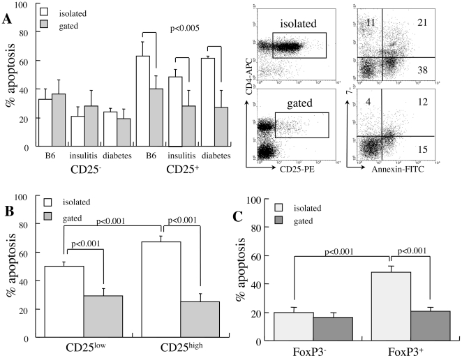
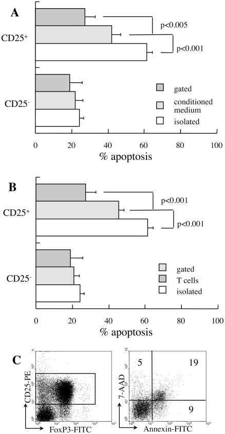
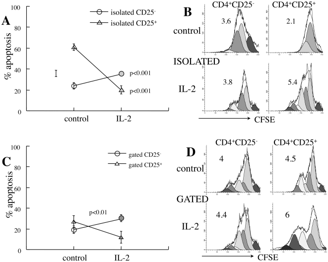
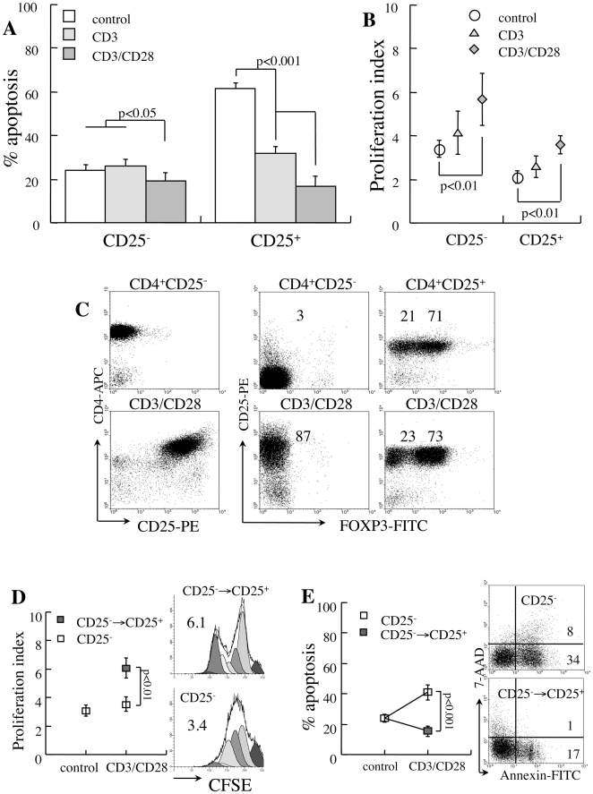
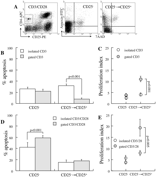
Principal findings: Apoptotic cells are short lived in vivo and death occurs primarily during isolation, manipulation and culture. Excessive susceptibility of CD25(+) T cells to spontaneous apoptosis is characteristic of isolated subsets, however disappears when death is measured in mixed splenocyte cultures. In variance, CD25(-) T cells display balanced sensitivity to apoptosis under both conditions. The isolation procedure removes soluble factors, IL-2 playing a significant role in sustaining Treg viability. In addition, pro- and anti-apoptotic signals are transduced by cell-to-cell interactions: CD3 and CD28 protect CD25(+) T cells from apoptosis, and in parallel sensitize naïve effector cells to apoptosis. Treg viability is modulated both by other T cells and other subsets within mixed splenocyte cultures. Variations in sensitivity to apoptosis are often hindered by fast proliferation of viable cells, therefore cycling rates are mandatory to adequate interpretation of cell death assays.

Conclusions: The sensitivity of purified Treg to apoptosis is dominated by cytokine deprivation and absence of cell-to-cell interactions, and deviate significantly from measurements in mixed populations. Balanced sensitivity of naïve/effector and regulatory T cells to apoptosis in NOD mice argues against the concept that differential susceptibility affects disease evolution and progression.


Conflict of interest statement

Competing Interests: The authors have declared that no competing interests exist.

Figures

Figure 1
Figure 1. Impact of cell isolation on sensitivity to apoptosis.
A. Apoptosis of CD4+CD25 and CD4+CD25+ subsets measured after 48 hours in isolated cell suspensions (n = 5) and by gating in mixed cultures (n = 5). Cells were harvested from wild type C57BL/6, prediabetic NOD females aged 14 weeks and new onset diabetic NOD mice. Representative measurements of 7-AAD and Annexin-V incorporation are shown for isolated and mixed populations (gated) from diabetic NOD females. B. Differential apoptosis in reference to low and high CD25 expression measured in isolated CD25+ subsets (n = 4) and by gating in mixed cultures (n = 5). C. Spontaneous apoptosis in reference to FoxP3 expression in the isolated CD25+ subset (n = 3) and in mixed cultures (n = 3).
Figure 2
Figure 2. Soluble and cellular factors affecting susceptibility to apoptosis.
A. CD25 and CD25+ T cells isolated from new onset diabetic NOD females were incubated for 48 hours in conditioned medium from CD25 T cells stimulated with surface-bound anti CD3 and anti-CD28 antibodies (n = 3). Data are compared to corresponding measurements of isolated cells and mixed cultures. B. Spontaneous apoptosis after 48 hours of culture of isolated CD25+ T cells (n = 4) and gated subsets in mixed cultures (n = 5) following B220, GR-1 and MAC-1 depletion (n = 4). C. Equal numbers of isolated CD25 and CD25+ T cells from diabetic NOD mice were mixed for determination of apoptosis after 48 hours of culture in the CD25+ subset (gate). Data are representative of four independent incubations.
Figure 3
Figure 3. Influence of IL-2 on CD4+ T cell apoptosis.
A. Apoptosis of CD4+CD25 (n = 5) and CD4+CD25+ T cells (n = 4) isolated from new onset diabetic NOD females and incubated for 48 hours with and without 2000 U/ml IL-2. B. Demonstrative measurements of proliferation rates of isolated CD25 and CD25+ cells as determined from CFSE dilution (representative of 4 independent measurements). C. Impact of exogenous IL-2 supplementation on apoptosis measured by gating on CD25 and CD25+ subsets in mixed cultures (n = 4). D. Demonstrative plots of CFSE dilution in gated CD4+ subsets within mixed cultures (representative of 4 measurements).
Figure 4
Figure 4. Sensitivity to apoptosis of isolated CD4+ T cells under CD3 and CD28 stimulation.
CD25 and CD25+ T cells isolated from new onset diabetic NOD mice were stimulated with beads conjugated to anti-CD3 and anti-CD28. A. Apoptosis during 48 hours of incubation under CD3 (n = 4) and CD3/CD28 stimulation (n = 4). B. Proliferation rates determined from CFSE dilution. C. Isolated CD25 T cells convert to express CD25 without FoxP3 priming during 48 hours of CD3/CD28 stimulation. Isolated CD25+ T cells sustain Cd25 and FoxP3 expression during Cd3/Cd28 stimulation. D. Proliferation rates of cells unresponsive to stimulation (CD25) and the subset that upregulates CD25 expression (CD25→CD25+) and representative plots of CFSE dilution. E. Fractional apoptosis of responsive and unresponsive CD25 T cells under CD3/CD28 stimulation (n = 4) and representative plots of apoptosis.
Figure 5
Figure 5. Apoptosis under CD3/CD28 stimulation.
A. Representative plots demonstrating upregulation of CD25 in response to CD3/CD28 stimulation in mixed splenocyte cultures. B. Spontaneous apoptosis in isolated (n = 4) and gated cells (n = 5) in reference to responsiveness (CD25 expression) to CD3-activation, assuming negligible contribution of naturally occurring CD25+ T cells in mixed cultures. C. Proliferation rates under CD3-activation of responsive (CD25→CD25+) and non-responsive (CD25) subsets in isolated CD25 T cells (n = 4) and in mixed cultures (gated, n = 5). D. Fractional apoptosis of responsive and non-responsive cells to CD3/CD28 stimulation in mixed cultures (n = 5). E. Proliferation rates under CD3/CD28 stimulation.

References

    1. Shevach EM. Regulatory T cells in autoimmmunity. Annu Rev Immunol. 2000;18:423–449. - PubMed
    1. Maloy KJ, Powrie F. Regulatory T cells in the control of immune pathology. Nat Immunol. 2001;2:816–822. - PubMed
    1. Bluestone JA, Abbas AK. Natural versus adaptive regulatory T cells. Nat Rev Immunol. 2003;3:253–257. - PubMed
    1. Sakaguchi S. Naturally arising CD4+ regulatory T cells for immunologic self-tolerance and negative control of immune responses. Annu Rev Immunol. 2004;22:531–562. - PubMed
    1. Sakaguchi S, Sakaguchi N, Asano M, Itoh M, Toda M. Immunologic self-tolerance maintained by activated T cells expressing IL-2 receptor α-chains (CD25). Breakdown of a single mechanism of self-tolerance causes various autoimmune diseases. J Immunol. 1995;155:1151–1164. - PubMed

Publication types