. 2010 Dec 29;5(12):e15690.
doi: 10.1371/journal.pone.0015690.

Oral methylthioadenosine administration attenuates fibrosis and chronic liver disease progression in Mdr2-/- mice

Affiliations

Oral methylthioadenosine administration attenuates fibrosis and chronic liver disease progression in Mdr2-/- mice

M Ujue Latasa et al. PLoS One. .

Abstract

Background: Inflammation and fibrogenesis are directly related to chronic liver disease progression, including hepatocellular carcinoma (HCC) development. Currently there are few therapeutic options available to inhibit liver fibrosis. We have evaluated the hepatoprotective and anti-fibrotic potential of orally-administered 5'-methylthioadenosine (MTA) in Mdr2(-/-) mice, a clinically relevant model of sclerosing cholangitis and spontaneous biliary fibrosis, followed at later stages by HCC development.

Methodology: MTA was administered daily by gavage to wild type and Mdr2(-/-) mice for three weeks. MTA anti-inflammatory and anti-fibrotic effects and potential mechanisms of action were examined in the liver of Mdr2(-/-) mice with ongoing fibrogenesis and in cultured liver fibrogenic cells (myofibroblasts).

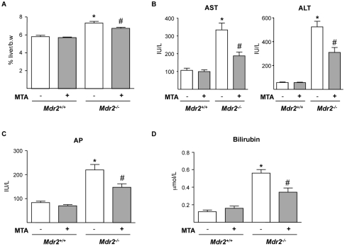
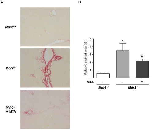
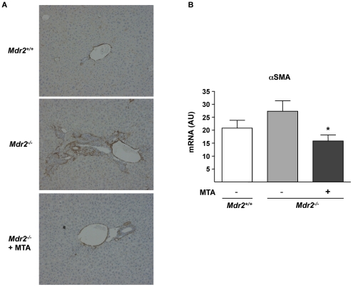
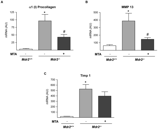
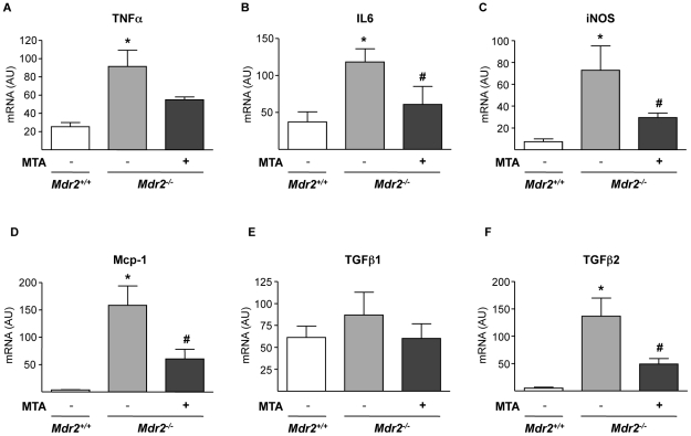
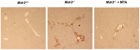
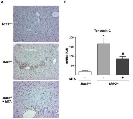
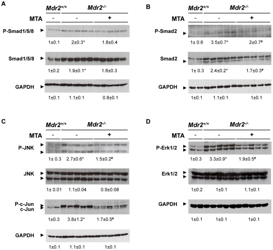
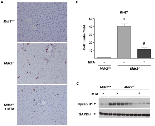
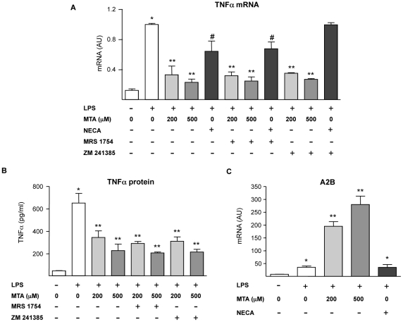
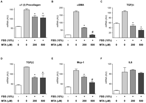
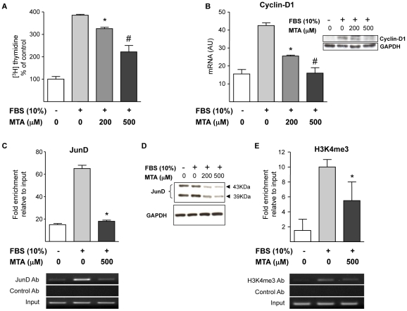
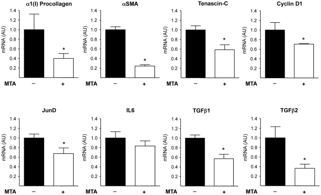
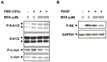
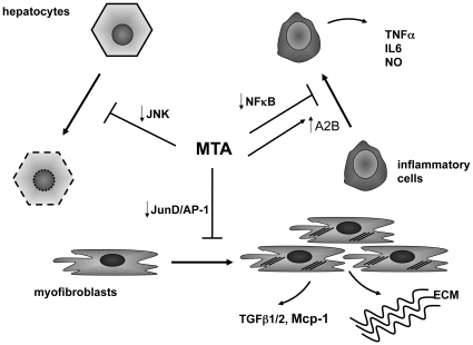
Principal findings: MTA treatment reduced hepatomegaly and liver injury. α-Smooth muscle actin immunoreactivity and collagen deposition were also significantly decreased. Inflammatory infiltrate, the expression of the cytokines IL6 and Mcp-1, pro-fibrogenic factors like TGFβ2 and tenascin-C, as well as pro-fibrogenic intracellular signalling pathways were reduced by MTA in vivo. MTA inhibited the activation and proliferation of isolated myofibroblasts and down-regulated cyclin D1 gene expression at the transcriptional level. The expression of JunD, a key transcription factor in liver fibrogenesis, was also reduced by MTA in activated myofibroblasts.

Conclusions/significance: Oral MTA administration was well tolerated and proved its efficacy in reducing liver inflammation and fibrosis. MTA may have multiple molecular and cellular targets. These include the inhibition of inflammatory and pro-fibrogenic cytokines, as well as the attenuation of myofibroblast activation and proliferation. Downregulation of JunD and cyclin D1 expression in myofibroblasts may be important regarding the mechanism of action of MTA. This compound could be a good candidate to be tested for the treatment of (biliary) liver fibrosis.


Conflict of interest statement

Competing Interests: The authors have declared that no competing interests exist.

Figures

Figure 1
Figure 1. Oral administration of MTA to Mdr2−/− mice reduces hepatomegaly and serum parameters of liver injury.
Mdr2+/+ and Mdr2−/− mice were treated with MTA during three weeks. Increased liver-to-body weight ratio (%) in Mdr2−/− mice was reduced by MTA (A). Serum transaminases (B), alkaline phosphatase (C) and bilirubin (D), levels that are increased in Mdr2−/− mice were attenuated by MTA. *P<0.05 vs untreated Mdr2+/+ mice, #P<0.05 vs untreated Mdr2−/− mice.
Figure 2
Figure 2. MTA administration reduces the spontaneous fibrosis that develops in Mdr2−/− mice.
Representative Sirius Red-stained liver sections from Mdr2+/+, control Mdr2−/− and MTA-treated Mdr2−/− mice (Mdr2−/− + MTA) (A). Fibrosis was quantified as function of mean percentage of stained area (B). *P<0.05 vs Mdr2+/+ mice, #P<0.05 vs untreated Mdr2−/− mice.
Figure 3
Figure 3. MTA administration reduces αSMA expression in Mdr2−/− mice.
Immunohistochemical staining of αSMA in representative liver sections from wild type, control Mdr2−/− mice, and MTA-treated Mdr2−/− mice (Mdr2−/− + MTA). The increased number of positive periductal myofibroblasts was reduced upon MTA administration (A). αSMA mRNA levels in Mdr2+/+, control Mdr2−/−, and MTA-treated Mdr2−/− mice (B). AU: arbitrary units. *P<0.05 vs untreated Mdr2−/− mice.
Figure 4
Figure 4. Fibrosis-related gene expression in Mdr2−/− mouse liver is attenuated by MTA treatment.
mRNA levels of α1(I) procollagen (A), MMP13 (B) and TIMP1 (C), were measured in the liver of Mdr2+/+, control Mdr2−/−, and MTA-treated Mdr2−/− mice. AU: arbitrary units. *P<0.05 vs Mdr2+/+ mice, #P<0.05 vs untreated Mdr2−/− mice.
Figure 5
Figure 5. Effect of MTA on the expression of inflammatory cytokines and iNOS in Mdr2−/− mouse liver.
mRNA levels of TNFα (A), IL6 (B), iNOS (C), Mcp-1 (D), TGFβ1 (E) and TGFβ2 (F) were measured in the liver of Mdr2+/+, control Mdr2−/−, and MTA-treated Mdr2−/− mice. *P<0.05 vs Mdr2+/+ mice, #P<0.05 vs untreated Mdr2−/− mice.
Figure 6
Figure 6. MTA reduces the recruitment of leukocytes to the portal fields of Mdr2−/− mice.
Representative staining for the pan-leukocyte marker CD45 showing increased accumulation of CD45 positive cells in the portal field of Mdr2−/− mice compared to WT animals. Treatment with MTA reduced CD45 positive leukocyte accumulation. Representative images are shown.
Figure 7
Figure 7. Expression of the fibrosis-associated protein tenascin-C, up-regulated in Mdr2−/− mice, is attenuated by MTA administration.
Immunohistochemistry of tenascin-C in representative liver sections shows increased staining around portal areas in Mdr2−/− mice and its downregulation by MTA (A). Tenascin-C mRNA levels in the liver of Mdr2+/+, control Mdr2−/−, and MTA-treated Mdr2−/− mice (B). *P<0.05 vs Mdr2+/+ mice, #P<0.05 vs untreated Mdr2−/− mice.
Figure 8
Figure 8. MTA modulates intracellular signalling pathways activated in Mdr2−/− mouse liver.
Phosphorylation levels of Smad1/5/8 (A), Smad2 (B), JNK and c-Jun (C) and Erk1/2 (D) were analyzed by western blotting in liver extracts from Mdr2+/+, control Mdr2−/−, and MTA-treated Mdr2−/− mice. *P<0.05 vs Mdr2+/+ mice, #P<0.05 vs untreated Mdr2−/− mice.
Figure 9
Figure 9. Increased hepatocellular proliferation in Mdr2−/− mice is attenuated by MTA administration.
Immunohistochemical staining of Ki-67 in liver sections from Mdr2+/+, control Mdr2−/−, and MTA-treated Mdr2−/− mice (Mdr2−/−+MTA)(A). Ki-67 positive hepatocytes were counted in 30 high-power fields per mouse, n = 5 per group (B). *P<0.01 vs Mdr2+/+ mice, #P<0.05 vs untreated Mdr2−/− mice. Western blot analysis of cyclin-D1 protein in liver extracts from Mdr2+/+ mice, untreated Mdr2 / and MTA-treated Mdr2−/− mice (C).
Figure 10
Figure 10. Effect of MTA on the activation of inflammatory cells: interaction with adenosine receptors.
As indicated in the figure serum-starved murine monocyte/macrophages RAW 264.7 cells were pre-treated for 30 min with the A2A receptor antagonist ZM 241385 (10 µM) or the A2B receptor antagonist MRS 1754 (1 µM), then MTA or the adenosine receptor agonist NECA (20 µM) where added to the cultures for another 30 min. Subsequently and where indicated cells were treated with LPS (50 ng/ml) for up to 5 h. TNFα mRNA levels in cell lysates (A) and TNFα protein contents in conditioned media (B) were measured. *P<0.01 vs control, **P<0.01 vs LPS, #P<0.05 vs LPS. The expression of the adenosine A2B receptor was measured in RAW 264.7 cells pre-treated with MTA or the adenosine agonist NECA (20 µM) for 30 min and then where indicated with LPS (50 ng/ml) for up to 5 h (C). *P<0.01 vs control, **P<0.01 vs LPS.
Figure 11
Figure 11. Effects of MTA on liver myofibroblast activation and cytokine expression.
Primary myofibroblasts were serum starved (12 h) and then treated (12 h) with 10% FCS and MTA as indicated. mRNA levels of α1(I)procollagen (A), αSMA (B), TGFβ1 (C), TGFβ2 (D), Mcp-1 (E) and IL6 (F) were measured at the end of treatments. *P<0.05 vs FCS, #P<0.05 vs FCS+MTA 200 µM.
Figure 12
Figure 12. MTA inhibits DNA synthesis in myofibroblasts.
MTA Effects on cyclin D1 and JunD expression. Effect of MTA on FCS-induced [3H]thymidine incorporation in myofibroblasts treated as indicated (24 h) (A). Cyclin D1 mRNA expression in myofibroblasts treated (12 h) with 10% FCS and MTA as indicated. Inset shows a Western blot analysis of cyclin D1 protein (B). ChIP assay measuring the binding of JunD to the −905 to −725 cyclin D1 promoter region encompassing a key AP-1 binding site in myofibroblasts treated as indicated (12 h). Immunoprecipitated genomic DNA corresponding to the −905 to −725 cyclin D1 promoter region was quantified by real time PCR as indicated in Methods section. A representative gel picture of amplified DNA fragments is shown (C). Western blot analysis of JunD protein levels in myofibroblasts treated as indicated for 6 h (D). ChIP assay measuring the binding of tri-methylated-H3K4 (H3K4me3) to the same cyclin-D1 promoter region in myofibroblasts treated as indicated (12 h). A representative gel picture of amplified DNA fragments is shown (E). *P<0.05 vs FCS, #P<0.05 vs FCS+MTA 200 µM.
Figure 13
Figure 13. Expression of fibrogenic activation and cellular proliferation-related genes in myofibroblasts isolated from the liver of control and MTA-treated Mdr2−/− mice.
Mdr2−/− mice were treated for three weeks with MTA or vehicle as described in the Methods section. At the end of the treatments hepatic myofibroblasts were isolated and the mRNA levels of the indicated genes were measured. Data are means ±SEM of three independent cell preparations per condition. *P<0.05 vs vehicle-treated Mdr2−/− mice.
Figure 14
Figure 14. Effect of MTA on liver myofibroblast intracellular signalling pathways.
Liver myofibroblasts were pre-treated with MTA for 30 min in the absence of serum and then stimulated with 10% FCS for 30 min or PDGF (20 ng/ml) for 60 min. Phosphorylation levels of Erk1/2 and c-Jun upon FCS stimulation (A), and S6 ribosomal protein after PDGF treatment (B) were analyzed by western blotting. Representative blots are shown.
Figure 15
Figure 15. Potential mechanisms of action of MTA in Mdr2−/− mice.
Our in vivo and in vitro observations indicate that MTA displays different actions that may underlie its beneficial effects on the course of liver injury and fibrosis in Mdr2−/− mice. MTA may exert a direct cytoprotective effect on hepatocytes by reducing JNK activity, preventing hepatocellular death and further inflammation. MTA also inhibits the production of cytokines by inflammatory cells, likely through interference with NFκB activity as described before –. In addition, enhanced expression of adenosine A2B receptors may contribute to the anti-inflammatory pharmacological profile of MTA. This compound can also exert direct effects on extracellular matrix (ECM) producing cells (myofibroblasts), reducing their activation, proliferation and the production of pro-fibrogenic factors. Inhibition of JunD expression may be an important event in the antifibrogenic action of MTA.

References

    1. Williams R. Global challenges in liver disease. Hepatology. 2006;44:521–6. - PubMed
    1. Siegel AB, Zhu AX. Metabolic syndrome and hepatocellular carcinoma. Cancer. 2009;115:5651–5661. - PMC - PubMed
    1. Bataller R, Brenner DA. Liver fibrosis. J Clin Invest. 2005;115:209–218. - PMC - PubMed
    1. Elsharkawy AM, Mann DA. Nuclear factor-κB and the hepatic inflammation-fibrosis-cancer axis. Hepatology. 2007;46:590–597. - PubMed
    1. Lotersztajn S, Julien B, Teixeira-Clerc F, Grenard P, Mallat A. Hepatic fibrosis: molecular mechanisms and drug targets. Annu Rev Pharmacol Toxicol. 2005;45:605–628. - PubMed

Publication types

MeSH terms