

Spontaneous biloma managed with endoscopic retrograde cholangiopancreatography and percutaneous drainage: a case report
- PMID: 21210994
- PMCID: PMC3023755
- DOI: 10.1186/1752-1947-5-3
Spontaneous biloma managed with endoscopic retrograde cholangiopancreatography and percutaneous drainage: a case report
Abstract
Introduction: Spontaneous biloma formation is a very rare condition, which mandates immediate treatment.
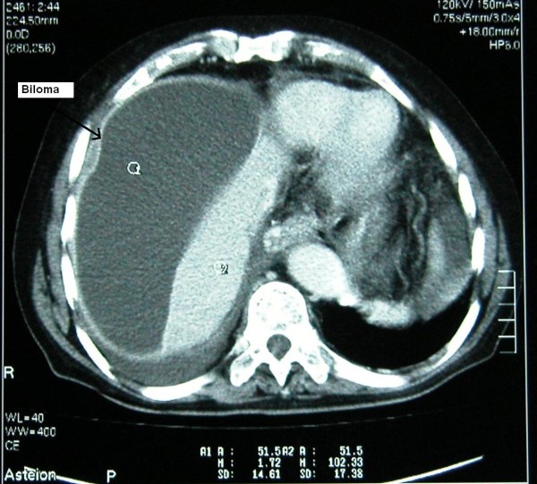
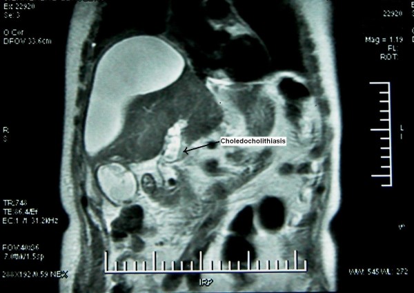
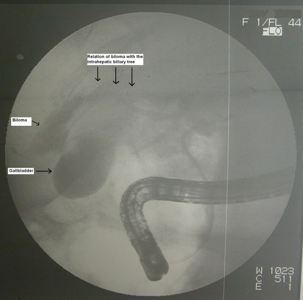
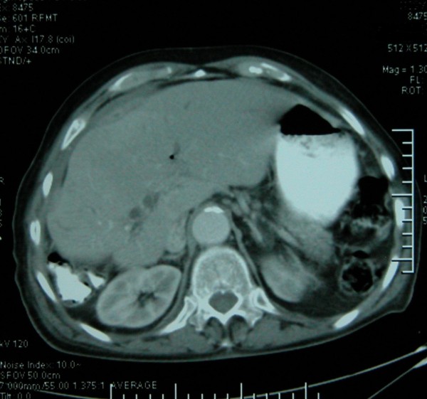
Case presentation: An 80-year-old Caucasian man was referred to our department with a diagnosis of intra-abdominal collection located in his right upper quadrant. Further radiological examination demonstrated multiple calculi in his gallbladder and common bile duct. Our patient underwent endoscopic retrograde cholangiopancreatography and the stones in the common bile duct were extracted. Percutaneous drainage of the abdominal collection revealed a spontaneous biloma formation. Continuous drainage of bile persisted for one week, so endoscopic retrograde cholangiopancreatography was repeated and a 10Fr stent was placed; subsequently the biliary leak ceased and our patient was discharged. A control abdominal computed tomography did not show any residual fluid collection.
Conclusion: Spontaneous biloma formation is a very rare incidence; awareness is necessary for prompt recognition and treatment.
Figures
References
-
- Gould L, Patel A. Ultrasound detection of extrahepatic encapsulated bile: "biloma". AJR Am J Roentgenol. 1979;132(6):1014–1015. - PubMed
-
- Vazquez JL, Thorsen MK, Dodds WJ, Quiroz FA, Martinez ML, Lawson TL, Stewart ET, Foley WD. Evaluation and treatment of intraabdominal bilomas. AJR Am J Roentgenol. 1985;144(5):933–938. - PubMed
-
- Fujiwara H, Yamamoto M, Takahashi M, Ishida H, Ohashi O, Onoyama H, Takeyama Y, Kuroda Y. Spontaneous rupture of an intrahepatic bile duct with biloma treated by percutaneous drainage and endoscopic sphincterotomy. Am J Gastroenterol. 1998;93(11):2282–2284. doi: 10.1111/j.1572-0241.1998.00636.x. - DOI - PubMed
LinkOut - more resources
Full Text Sources
