. 2011 Jan 7;88(1):106-14.
doi: 10.1016/j.ajhg.2010.12.004.

NEK1 mutations cause short-rib polydactyly syndrome type majewski

Affiliations

NEK1 mutations cause short-rib polydactyly syndrome type majewski

Christian Thiel et al. Am J Hum Genet. .

Abstract

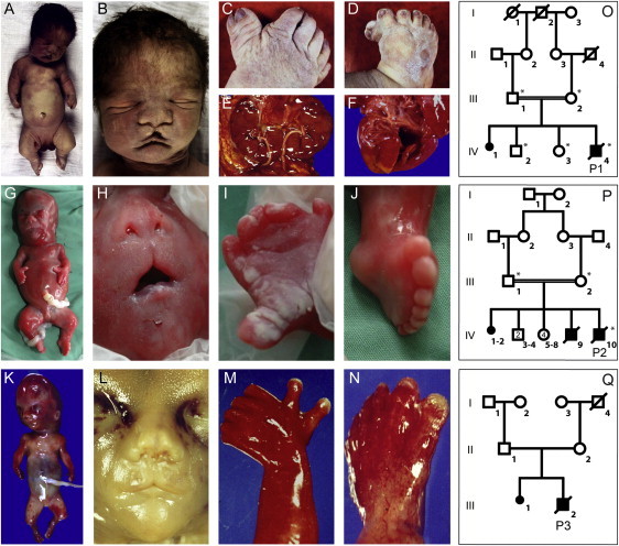
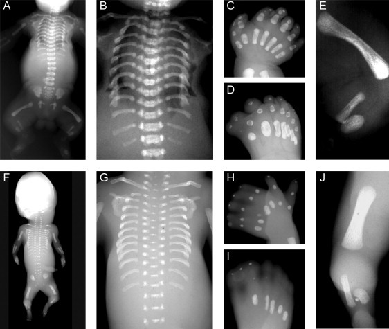
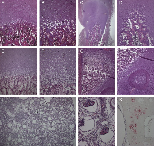
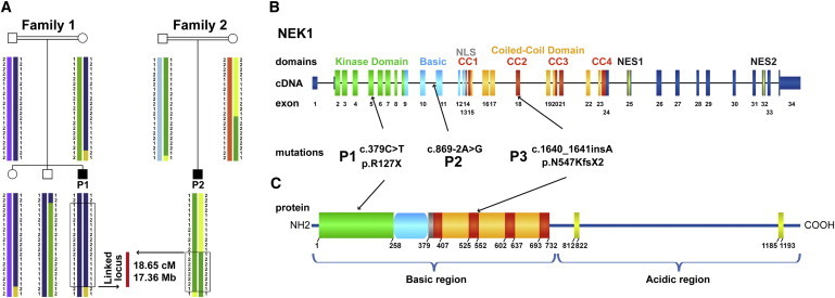
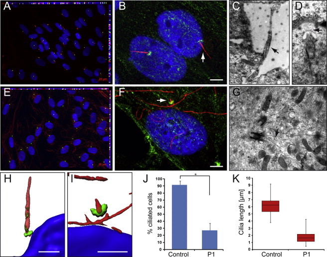
Defects of ciliogenesis have been implicated in a wide range of human phenotypes and play a crucial role in signal transduction and cell-cycle coordination. We used homozygosity mapping in two families with autosomal-recessive short-rib polydactyly syndrome Majewski type to identify mutations in NEK1 as an underlying cause of this lethal osteochondrodysplasia. NEK1 encodes a serine/threonine kinase with proposed function in DNA double-strand repair, neuronal development, and coordination of cell-cycle-associated ciliogenesis. We found that absence of functional full-length NEK1 severely reduces cilia number and alters ciliar morphology in vivo. We further substantiate a proposed digenic diallelic inheritance of ciliopathies by the identification of heterozygous mutations in NEK1 and DYNC2H1 in an additional family. Notably, these findings not only increase the broad spectrum of ciliar disorders, but suggest a correlation between the degree of defective microtubule or centriole elongation and organization and the severity of the resulting phenotype.


Figures

Figure 1
Figure 1
Morphological Features of the Investigated Patients with SRPS Type Majewski (A–F) The affected individual of the consanguineous family 1 was born at 36 wk of gestation and deceased 1 hr postpartum. Clinical evaluation showed severe extreme shortening of the ribs with a narrow thorax, a bulging forehead, depressed nasal bridge, and median cleft (B); extreme pre- and postaxial polysyndactyly with seven fingers and toes (C, D); horseshoe configuration with fused lower parts of both kidneys (E); and a small ventricular septal defect and open foramen ovale of the heart (F). (G–N) Similar clinical features were observed in the affected individual of family 2 (G–J) at 21 wk of gestation and the affected individual of family 3 (K–N) at 19 wk of gestation, with hydropic fetus, flat nose and median cleft, high and broad forehead, and mild microretrognathia, as well as pre- and postaxial polysyndactyly in both hands and feet. (O–Q) Pedigrees of the investigated family 1 (O), family 2 (P), and family 3 (Q). Individuals marked with asterisks were included in the linkage analysis.
Figure 2
Figure 2
Radiographic Features of the Investigated Patients with SRPS Type Majewski Both affected individuals, from family 1 (A–E) and family 3 (F–J), presented with severely shortened and horizontal ribs (A, B, F, G), squared scapulae and elevated clavicles with lateral kinking (A, F), normal vertebral column and pelvis and regular metaphyseal margins of the tubular bones, incompletely calcified and irregularly shaped metacarpal and metatarsal bones (C, D, H, I), shortened humerus and femurs, and extreme shortening of the tibial bones.
Figure 3
Figure 3
Histological and Immunohistochemical Analysis (A, B, E, F) Normal resting cartilage and growth plates with disorganization of endochondral ossification in the affected individuals P1 (A, B) and P3 (E, F). Note the reduced number of chondrocytes in the epiphyseal zone of proliferation and the diminished cartilage-column formation of the growth zone. (C, D) Additional lack of columnization of the enchondral ossification of the epiphyses of the ribs. Here, the growth zone was lined in a semicircle around the osseous center of the rib bone. (G, H) The growing cartilage of the tibial bone formed a semicircle around the metadiaphyseal zone, with heterotopic ossification. (I, J) Polycystic kidney disease was found in affected individual P1 with dilatated collecting tubules, glomerules of differing size, and well developed glomerules with dense convolutes of capillary vessels. (K) Immunohistochemical analysis showed small islands of persistent hypertrophic chondrocytes with expression of collagen X within osseous trabecules.
Figure 4
Figure 4
Identification of NEK1 Mutations as Underlying Cause of SRPS Majewski Type by Positional Cloning (A) Haplotype structure analysis in the two consanguineous families 1 and 2 refined a locus to 18.65 cM / 17.36 Mb by homozygosity mapping including 54 RefSeq and hypothetical genes on chromosome 4q32.1-q34.3. (B) Sanger sequencing of the prioritized functional candidate gene NEK1 revealed a homozygous nonsense mutation in P1 (p.R127X), a homozygous splice site mutation in P2 (c.869-2A>G), and a heterozygous insertion in P3 (c.1640_1641_insA). (B, C) Schematic drawing of domain-coding exons according to the prediction of the domain structure in mice and protein mice-human homology. Abbreviations are as follows: kinase domain, N-terminal kinase domain (green); basic, basic domain (light blue); NLS, nuclear localization signal (gray); CC1-4, coiled-coil domains (red and orange); NES1-2, nuclear export sequence (yellow).
Figure 5
Figure 5
SRPS Majewski Type Fibroblasts Have a Severely Reduced Number and Structurally Abnormal Cilia The cells were grown on coverslips or on cell-culture dishes with gas-permeable base to confluence and then serum starved for 5 days, fixed, and treated for immunofluorescence analyses or TEM. (A, E) Immunofluorescence for acetylated α-tubulin (red), pericentrin (green), and DAPI (blue) shows normal presentation of cilia in control fibroblast cells (A), but missing or severely shortened cilia with normal appearance of the basal body and increased cytoplasmatic acetylated α-tubulin in P1 fibroblast cells (E). (J) Only 27% of P1 cells have verifiable cilia, in contrast to 92% of control cells (t test, p value 4 × 10−10). (B, F) Immunofluorescence for α-tubulin (red), NEK1 (green), and DAPI (blue) suggests the presence of the 63 kDa isoform NEK1 seen by immunoblotting in the PCM in control and P1 cells (white bar represents 5μm). The latter indicates presence of a physiological NEK1 isoform not affected by the nonsense mutation. Such an isoform lacking the N-terminal region would not be able to maintain cilial formation because of lack of the kinase domain. (K) Cilia length was measured at 6.2 ± 1.2 μm on average (median 6.2 μm, 25th centile 5.3 μm, 75th centile 6.8 μm) in control cells but only 1.7 ± 0.7 μm (median 1.6 μm, 25th centile 1.2 μm, 75th centile 2.2 μm) in P1 fibroblast cells (t test, p value 1.7 × 10−69). (C, D, G) Electron micrographs illustrating representative images from normal control fibroblast cells (C, D) and P1 fibroblast cells (F). (C) shows a primary cilia in full length, whereas in (D) the TEM image illustrates a primary cilia at the beginning of ciliogenesis (stage 2). In contrast, in P1 fibroblast cells (G), we found only centrioles at stage 1 of ciliogenesis with an abnormal microtubule growth (arrow) but no enveloping ciliary pocket around. (H) Imaris three-dimensional construction of normal cilia reveals the location of NEK1 at the basal body and within the cilium, suggesting participation in the intraflagellular transport. (I) In contrast, P1 cell cilia are shortened, with a broad base and a thin tip.

References

    1. Rohatgi R., Milenkovic L., Scott M.P. Patched1 regulates hedgehog signaling at the primary cilium. Science. 2007;317:372–376. - PubMed
    1. D'Angelo A., Franco B. The dynamic cilium in human diseases. Pathogenetics. 2009;2:3. - PMC - PubMed
    1. Satir P., Christensen S.T. Overview of structure and function of mammalian cilia. Annu. Rev. Physiol. 2007;69:377–400. - PubMed
    1. Rosenbaum J.L., Witman G.B. Intraflagellar transport. Nat. Rev. Mol. Cell Biol. 2002;3:813–825. - PubMed
    1. Cole D.G., Snell W.J. SnapShot: Intraflagellar transport. Cell. 2009;137:784. - PubMed

Publication types

MeSH terms