. 2011 Jan 7;88(1):115-20.
doi: 10.1016/j.ajhg.2010.12.006.

Mutations in the 5' UTR of ANKRD26, the ankirin repeat domain 26 gene, cause an autosomal-dominant form of inherited thrombocytopenia, THC2

Affiliations

Mutations in the 5' UTR of ANKRD26, the ankirin repeat domain 26 gene, cause an autosomal-dominant form of inherited thrombocytopenia, THC2

Tommaso Pippucci et al. Am J Hum Genet. .

Abstract

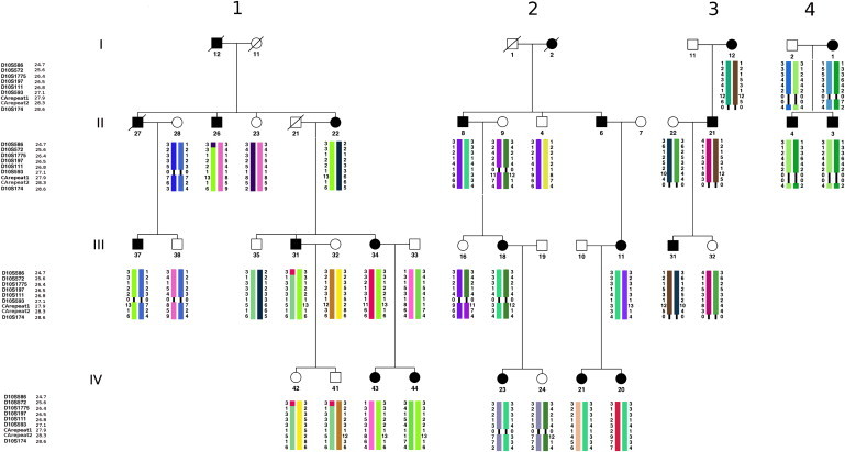
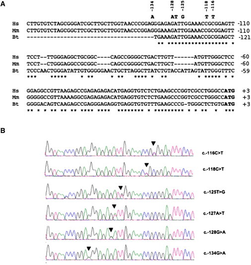
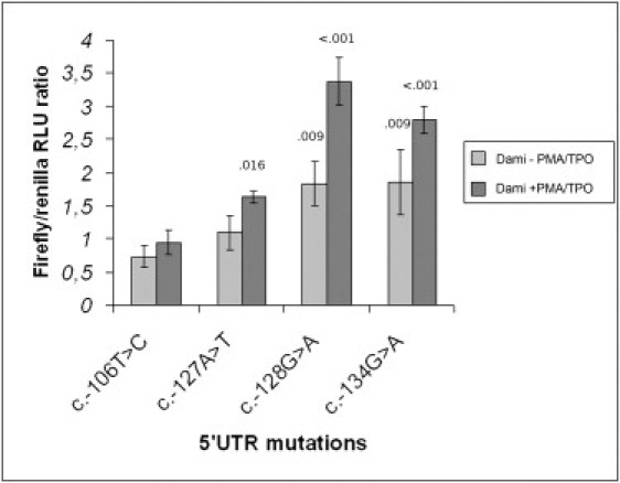
THC2, an autosomal-dominant thrombocytopenia described so far in only two families, has been ascribed to mutations in MASTL or ACBD5. Here, we show that ANKRD26, another gene within the THC2 locus, and neither MASTL nor ACBD5, is mutated in eight unrelated families. ANKRD26 was also found to be mutated in the family previously reported to have an ACBD5 mutation. We identified six different ANKRD26 mutations, which were clustered in a highly conserved 19 bp sequence located in the 5' untranslated region. Mutations were not detected in 500 controls and are absent from the 1000 Genomes database. Available data from an animal model and Dr. Watson's genome give evidence against haploinsufficiency as the pathogenetic mechanism for ANKRD26-mediated thrombocytopenia. The luciferase reporter assay suggests that these 5' UTR mutations might enhance ANKRD26 expression. ANKRD26 is the ancestor of a family of primate-specific genes termed POTE, which have been recently identified as a family of proapoptotic proteins. Dysregulation of apoptosis might therefore be the pathogenetic mechanism, as demonstrated for another thrombocytopenia, THC4. Further investigation is needed to provide evidence supporting this hypothesis.


Figures

Figure 1
Figure 1
Linkage to Chromosome 10p11.1-p12 in Four THC2 Families Segregation of microsatellite marker haplotypes in the THC2 locus on chromosome 10p11.1-p12 (with the corresponding Mb positions) in the two large THC2-linked families (Family 1 and 2) and in the two smaller families (families 3 and 4). Black symbols indicate affected individuals, and white symbols indicate healthy ones. Slashed symbols mean that those individuals are deceased. Only individuals for whom the corresponding haplotypes are reported were genotyped. Families 1 and 2, which carry the c.-128G>A and c.-127A>T mutations, respectively (Table 1), are consistent with linkage at the THC2 locus. Families 3 and 4 do not provide significant LOD scores, but their 10p11.1-p12 region segregates consistently with the disease. Family 3 carries the c.-118C>T mutation (Table 1). No mutation in ANKRD26 was found in the affected members of family 4, and therefore segregation of the haplotype is probably not related to the disease in this family. A 0/0 in the haplotype means unsuccessful genotyping for the marker in that individual. Haplotype representation was obtained with Haplopainter version 1.0.
Figure 2
Figure 2
The 5′-UTR-Mutated Sequences of ANKRD26 (A) Alignment of the 5′ UTRs of orthologs from Homo sapiens (Hs: NM_014915.2), Macaca mulatta (Mm: XM_002808496.1), and Bos taurus (Bt: NM_001113767.1). Nucleotide changes are in bold. Stars indicate matching sites. (B) Electropherograms showing the six different heterozygous mutations identified in THC2 families (Table 1) .
Figure 3
Figure 3
Firefly/Renilla RLU Ratios, Normalized against Wild-Type, for Each of the 5′ UTR Variants in the Functional Study Scale bars represent means ± standard deviation. Corresponding values of each variant for – PMA/TPO Dami cells (light gray) and + PMA/TPO Dami cells (dark gray) are as follows: c.-106T>C—0.73 ± 0.16, 0.94 ± 0.19; c.-127A>T— 1.09 ± 0.26, 1.63 ± 0.91; c.-128G>A—1.83 ± 0.19, 3.37 ± 0.36; and c.-134G>A—1.84 ± 0.49, 2.79 ± 0.20. The assumption of homogeneity of variances was respected both in −PMA/TPO and in +PMA/TPO Dami (Levene's statistic > .13 and .36, respectively). Normality of the distribution was respected for all the samples (Kolmogorov-Smirnov Z Test). ANOVA rejected the null hypothesis of equality of means in both groups. Significant p values at the 5% level for a Dunnett's test against the c.-106T>C control are reported above the corresponding column of the histogram.

References

    1. Nurden A.T., Nurden P. Inherited thrombocytopenias. Haematologica. 2007;92:1158–1164. - PubMed
    1. Balduini C.L., Savoia A. Inherited thrombocytopenias: Molecular mechanisms. Semin. Thromb. Hemost. 2004;30:513–523. - PubMed
    1. Noris P., Pecci A., Di Bari F., Di Stazio M.T., Di Pumpo M., Ceresa I.F., Arezzi N., Ambaglio C., Savoia A., Balduini C.L. Application of a diagnostic algorithm for inherited thrombocytopenias to 46 consecutive patients. Haematologica. 2004;89:1219–1225. - PubMed
    1. Savoia A., Del Vecchio M., Totaro A., Perrotta S., Amendola G., Moretti A., Zelante L., Iolascon A. An autosomal dominant thrombocytopenia gene maps to chromosomal region 10p. Am. J. Hum. Genet. 1999;65:1401–1405. - PMC - PubMed
    1. Drachman J.G., Jarvik G.P., Mehaffey M.G. Autosomal dominant thrombocytopenia: Incomplete megakaryocyte differentiation and linkage to human chromosome 10. Blood. 2000;96:118–125. - PubMed

Publication types

Supplementary concepts