Review
. 2011 Jan 7;41(1):8-19.
doi: 10.1016/j.molcel.2010.12.020.

Proteasome activators

Affiliations
Review

Proteasome activators

Beth M Stadtmueller et al. Mol Cell. .

Abstract

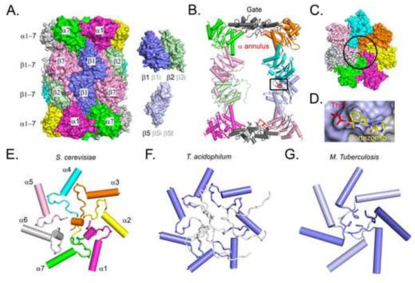
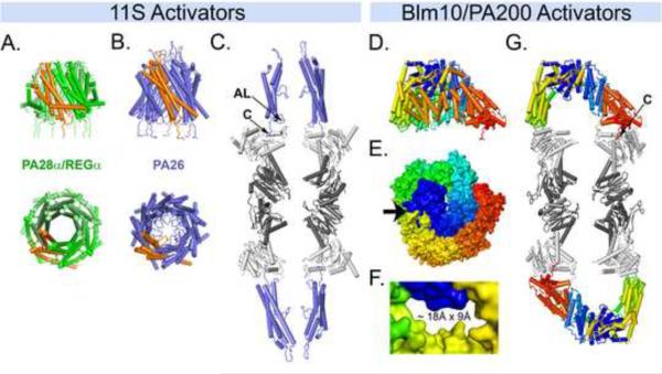
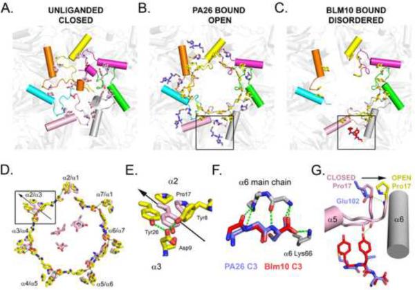
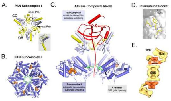
Proteasomes degrade a multitude of protein substrates in the cytosol and nucleus, and thereby are essential for many aspects of cellular function. Because the proteolytic sites are sequestered in a closed barrel-shaped structure, activators are required to facilitate substrate access. Structural and biochemical studies of two activator families, 11S and Blm10, have provided insights to proteasome activation mechanisms, although the biological functions of these factors remain obscure. Recent advances have improved our understanding of the third activator family, including the 19S activator, which targets polyubiquitylated proteins for degradation. Here we present a structural perspective on how proteasomes are activated and how substrates are delivered to the proteolytic sites.


Figures

Figure 1
Figure 1. Structure of the 20S proteasome
(A) Surface representation of the S.cerevisiae 20S proteasome (Groll et al., 1997). Individual subunits are labeled. Subunits to the right indicate that alternative counterparts can be expressed for some of the subunits. The bovine liver proteasome is closely superimposible to this structure (Unno et al., 2002). Archaeal and eubacterial 20S proteasomes are very similar but are comprised of one type of α and one type of β subunit, and are therefore seven-fold symmetric. (B) Cutaway side view cartoon representation. The region of the active site is indicated with a box. The gate region is colored gray. The α annulus, just interior from the gate, is an opening formed by loops (red) in the α subunits. (C) Top view of the S.cerevisiae 20S proteasome. The gate region is indicated with a circle. (D) Close-up of the β5 active site in complex with the inhibitor bortezomib. Corresponds to the boxed region in (B) (E) Top view cartoon of S.cerevisiae helix H0 and gating residues. This eukaryotic gate is sealed primarily by α2, α3, and α4, whose N-terminal residues are well ordered and participate in numerous hydrogen bonds and van der Waals contacts. Corresponds to the region circled in (C). (F) Same as (E) for an archaeal gate (Religa et al., 2010). Residues shown in white are highly mobile. (G) Same as (E) for a eubacterial gate (Li et al., 2010). This gate is ordered, but is quite different from the eukaryotic structure. The seven subunits are chemically identical but adopt a total of three different conformations at their N-termini, as indicated by the different shades.
Figure 2
Figure 2. ATP-Independent Activators
(A) Side and top views of PA28α/REGα in cartoon representation. One subunit is colored orange. (B) Same as (A) for PA26 as seen in proteasome complexes. Note the diaphragm-like structure formed in the central channel of PA26. (C) Cutaway of the PA26-S.cerevisiae proteasome complex. This figure was generated by removing subunits, to leave a total of eight proteasome and four PA26 subunits. The PA26 activation loop (AL) and C-terminus (C) are indicated for one of the subunits. (D) Side view of Blm10 as seen in the proteasome complex rainbow colored blue to red from N to C-termini. (E) Blm10 top view surface representation. Arrow indicates the largest opening through the Blm10 dome, which is not visible in this view. (F) Close-up of the Blm10 pore. (G) Cutaway of the Blm10-proteasome complex. The Blm10 C-terminus is indicated.
Figure 3
Figure 3. Mechanism of gate opening
(A) Top view of the S.cerevisiae proteasome gate in the closed conformation. The side chains of Tyr8, Asp9, Pro17, and Tyr26 (T.acidophilum numbering) of each subunit are colored pink. These residues stabilize the open conformation, and in some cases also make interactions that stabilize the closed conformation shown here. (B) Same as (A) for the PA26 complex, with Tyr8, Asp9, Pro17, and Tyr26 colored yellow. The C-terminal three residues of PA26 (blue) are shown in the four S.cerevisiae proteasome pockets where they are visible in the crystal structure (Forster et al., 2005). The pocket between α5 and α6 is boxed to encompass the PA26 C-terminal residues and also the Glu102 activation loop residue, which lies closer to the pseudo seven-fold axis and is shown for all seven PA26 subunits. The same open conformation is induced in an archaeal proteasome, when PA26 C-terminal residues bind equivalently to all seven pockets. (C) Same as (B) for the Blm10 complex. Blm10 makes extensive contacts that completely surround the proteasome entrance pore, and the C-terminal residues (red) bind in the α5/α6 pocket indicated. The Tyr8, Asp9, Pro17, and other N-terminal residues of some subunits that are ordered in the absence of an activator and become disordered upon binding Blm10 are shown in white in the position they occupy in the unliganded closed conformation. (D) Superposition of the Tyr8, Asp9, Pro17, and Tyr26 residues of the open (yellow) and closed (pink) gate conformations following overlap on the β subunits. This movement destabilizes packing of the N-terminal residues in the closed conformation and makes the pore wider so that a belt of Tyr8, Asp9 residues can assemble. The α2/α3 cluster, which undergoes the larges (3.5Å) displacement of a Pro17 residue, is boxed, and the outward direction of displacement upon opening is indicated with an arrow. The Pro17 residues of some α subunits are almost unchanged upon gate opening. (E) Close up of the α2/α3 cluster boxed in (D). (F) Close up of some interactions in the pocket boxed in (B) and (C) with PA26 and Blm10 superimposed. Main chain groups of PA26 and Blm10 C-terminal residues bind equivalently through Hydrogen bonds (dashed lines). (G) Superposition of the PA26 and Blm10 complexes in the α5/α6 pocket illustrating the different mechanisms of displacing Pro17. PA26 displaces Pro17 (in all seven subunits) by contacting the activation loop residue Glu102. Blm10 stabilizes the same Pro17 displacement (just of α5) by contacts of its penultimate tyrosine side chain.
Figure 4
Figure 4. ATP-dependent activators
(A) Top view of the N-terminal domains of PAN. The six subunits have identical sequences, but adopt alternating cis/trans conformations of Pro91 that allow formation of a trimer of coiled coils above a hexamer of OB domains. Inset shows a superposition of yellow (trans) and gray (cis) subunits on their OB domains to illustrate how the different conformations result in very different orientations for the N-terminal helix. (B) Top view of the C-terminal domain of PAN. The monomer crystal structure was determined and is modeled here on the hexameric structure of HslU, following the approach of (Zhang et al., 2009a). ATP/ADP sites are indicated in orange. Pore loop residues are colored green. (C) Cutaway side view of a composite model of the PAN hexamer based on the available domain structures. A substrate (red) is shown interacting with the N-terminal coiled-coils, which promote protein unfolding, and with a flexible segment extending through the conduit of OB domains to reach the pore loops that drive translocation. (D) Electron microscopic reconstruction of Drosophila 26S proteasome, adapted from (Nickell et al., 2009) with permission. Lid and base subcomplexes of the 19S activator are indicated. The volume assigned to the ATPases, as in the composite model of (C), is colored orange. (E) Model of Mycobacterium tuberculosis Pup-Mpa interactions (Wang et al., 2010). Pup binds the coiled-coil to present a disordered N-terminal segment that can traverse the double OB domain conduit of eubacterial ATPases, with both Pup and conjugated substrate being subsequently dragged into the proteasome by the ATPase translocation activity. Binding stoichiometry and packing considerations suggests the arrangement shown here, with just one Pup-substrate associated with the Mpa hexamer.

References

    1. Babbitt SE, Kiss A, Deffenbaugh AE, Chang YH, Bailly E, Erdjument-Bromage H, Tempst P, Buranda T, Sklar LA, Baumler J, et al. ATP hydrolysis-dependent disassembly of the 26S proteasome is part of the catalytic cycle. Cell. 2005;121:553–565. - PubMed
    1. Bajorek M, Finley D, Glickman MH. Proteasome disassembly and downregulation is correlated with viability during stationary phase. Curr Biol. 2003;13:1140–1144. - PubMed
    1. Baugh JM, Viktorova EG, Pilipenko EV. Proteasomes can degrade a significant proportion of cellular proteins independent of ubiquitination. J Mol Biol. 2009;386:814–827. - PMC - PubMed
    1. Bech-Otschir D, Helfrich A, Enenkel C, Consiglieri G, Seeger M, Holzhutter HG, Dahlmann B, Kloetzel PM. Polyubiquitin substrates allosterically activate their own degradation by the 26S proteasome. Nat Struct Mol Biol. 2009;16:219–225. - PubMed
    1. Benaroudj N, Zwickl P, Seemuller E, Baumeister W, Goldberg AL. ATP hydrolysis by the proteasome regulatory complex PAN serves multiple functions in protein degradation. Mol Cell. 2003;11:69–78. - PubMed

Publication types

MeSH terms

LinkOut - more resources