. 2011 Jan 7;41(1):67-81.
doi: 10.1016/j.molcel.2010.12.016.

Chromodomain-mediated oligomerization of HP1 suggests a nucleosome-bridging mechanism for heterochromatin assembly

Affiliations

Chromodomain-mediated oligomerization of HP1 suggests a nucleosome-bridging mechanism for heterochromatin assembly

Daniele Canzio et al. Mol Cell. .

Abstract

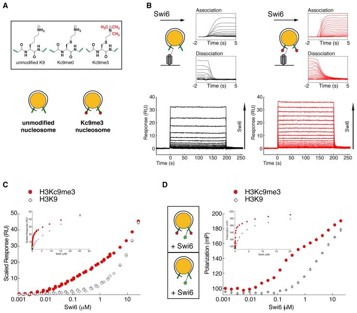
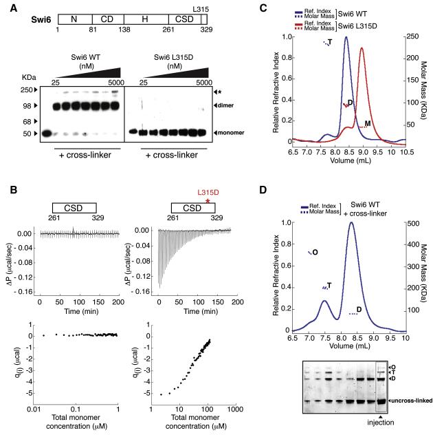
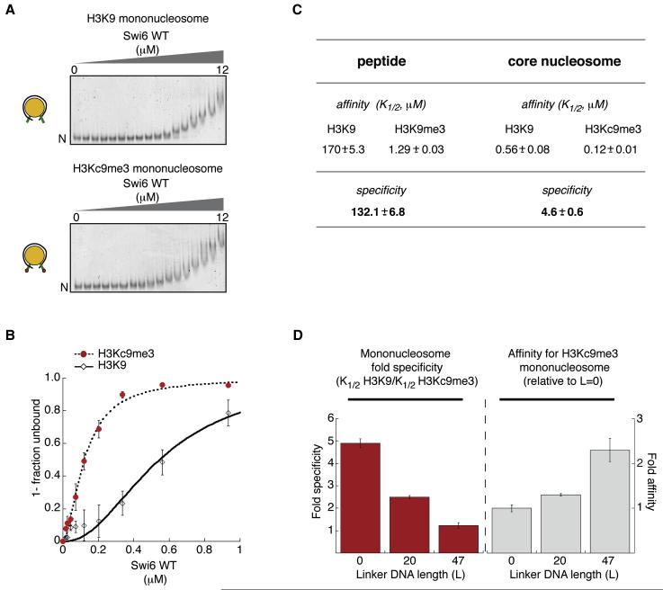
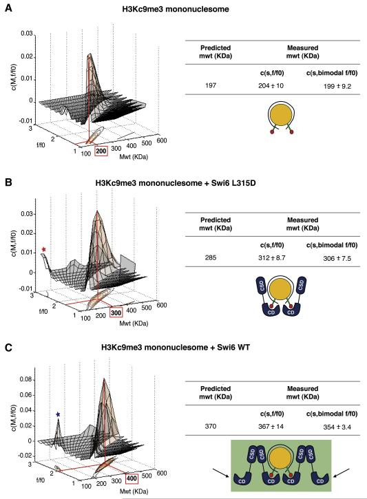
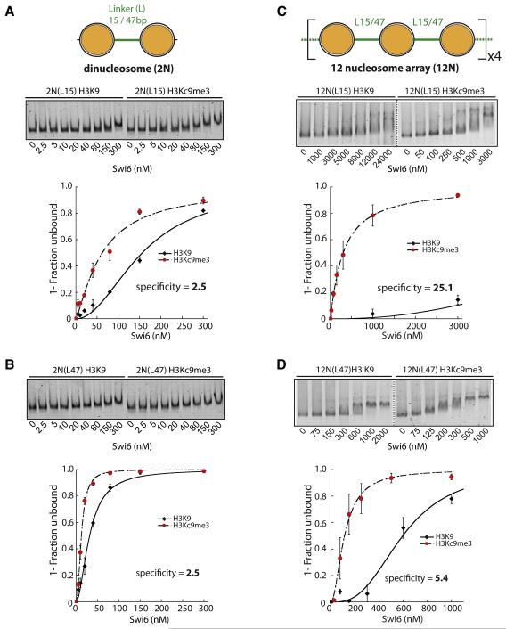
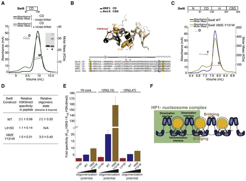
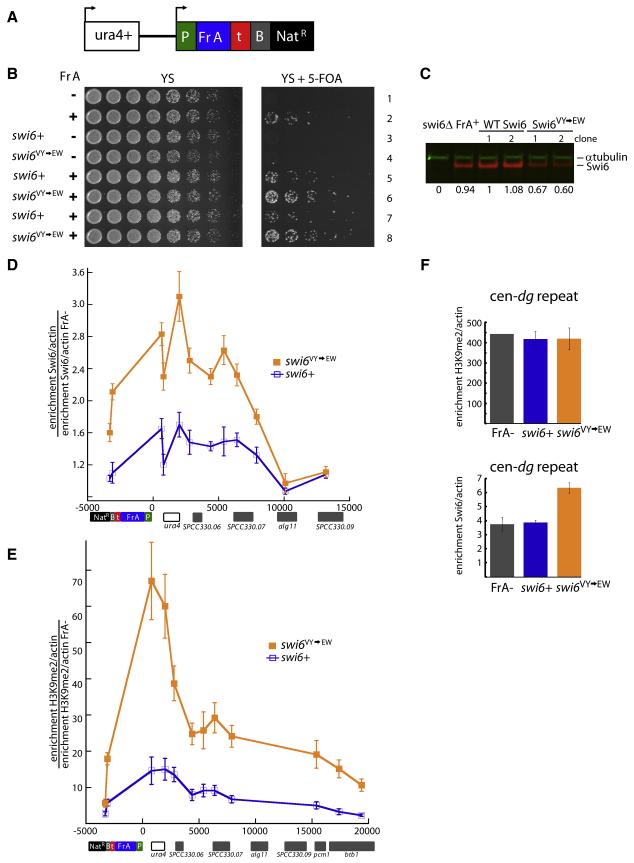
HP1 proteins are central to the assembly and spread of heterochromatin containing histone H3K9 methylation. The chromodomain (CD) of HP1 proteins specifically recognizes the methyl mark on H3 peptides, but the same extent of specificity is not observed within chromatin. The chromoshadow domain of HP1 proteins promotes homodimerization, but this alone cannot explain heterochromatin spread. Using the S. pombe HP1 protein, Swi6, we show that recognition of H3K9-methylated chromatin in vitro relies on an interface between two CDs. This interaction causes Swi6 to tetramerize on a nucleosome, generating two vacant CD sticky ends. On nucleosomal arrays, methyl mark recognition is highly sensitive to internucleosomal distance, suggesting that the CD sticky ends bridge nearby methylated nucleosomes. Strengthening the CD-CD interaction enhances silencing and heterochromatin spread in vivo. Our findings suggest that recognition of methylated nucleosomes and HP1 spread on chromatin are structurally coupled and imply that methylation and nucleosome arrangement synergistically regulate HP1 function.


Figures

Figure 1
Figure 1. Swi6 recognizes the H3K9 methyl mark within mononucleosomes and forms oligomers on mononucleosomes
(A) Schematics of the unmodified K9 and methyl lysine analog (MLA) Kc9me0 and Kc9me3 substrates (top panel) and of the unmodified (H3K9) and MLA methylated (H3Kc9me3) mononucleosomes assembled on the 147 bp 601 sequence (bottom). (B) Bottom Panels: Representative dose responses for H3K9 (black) and H3Kc9me3 (red) mononucleosomes. Schematic: H3K9 and H3Kc9me3 mononucleosomes captured on a streptavidin derivatized SPR chip. Top panels: Close up of the kinetics of association and dissociation. (C) Scaled isotherms for three independent dose responses of Swi6 against H3K9 (open diamonds) and H3Kc9me3 (filled circles) mononucleosome surfaces plotted on a semi-log scale. Plotted points represent the response at equilibrium, determined by averaging the signal over the final ten seconds of the sample injection. Inset: isotherms plotted on a linear scale. (D) Schematics: Mononucleosomes with fluorescein (green star) attached by a flexible linker at one end of the 147 bp DNA template. Average of three independent fluorescent polarization experiments for H3K9 (open diamonds) and H3Kc9me3 (filled circles) mononucleosomes are shown. Error bars represent s.e.m. All Swi6 concentrations represent monomer concentrations.
Figure 2
Figure 2. Swi6 forms distinct oligomeric states in the absence of chromatin
(A) Wild-type Swi6 (schematic on top) is largely a pre-formed dimer at low nM concentrations. Swi6 WT (left) and L315D (right) were treated at indicated concentrations with EDC and NHS cross-linkers. Treated proteins were separated by SDS-PAGE and detected by anti-FLAG western. Swi6 concentrations: uncross-linked 50 nM, cross-linked 25, 50, 100, 200, 500, 1000 and 5000 nM. (B) The CSD-CSD dimerization Kd < 17 nM. Top: Representative ITC thermogram profiles for the dissociation of WT CSD dimer (left) and L315D CSD dimer (right) at 15°C. Bottom: Graphs represent the respective binding isotherms plotted as heat changes per injection (qi) vs. total monomer concentration. (C) Multiangle light scattering (MALS) measurements for 20 μM WT Swi6 (blue) and 20 μM L315D mutant (red). Relative refractive index signals (solid lines, left y-axis) and derived molar masses (dotted lines, right y-axis) shown as a function of the elution volume. M: monomer, D: dimer, T: tetramer. (D) Top panel: Higher order oligomeric species of Swi6 stabilized by cross-linking. MALS measurements conducted as in (A). M: monomer, D: dimer, T: tetramer, O: octamer. Bottom panel: Aliquots of fractions collected from chromatography in (Top) were separated by denaturing SDS-PAGE and visualized by Sypro Red staining. The distribution of distinct oligomeric states thus visualized directly correlates with the oligomeric masses observed by MALS, while the presence of un-cross-linked Swi6 demonstrates Swi6 is not over cross-linked. All Swi6 concentrations represent monomer concentrations.
Figure 3
Figure 3. Swi6 displays lower specificity for the H3K9me3 mark in mononucleosomes compared to that in H3 tail peptides
(A) Representative gel shift using H3K9 or H3Kc9me3 mononucleosomes. Swi6 concentrations vary from 0 to 12 μM (0.6 fold dilutions). Unbound nucleosomes (N). (B) Quantification of three gel shift experiments using H3K9 (open diamonds) and H3Kc9me3 (filled circles) to determine K1/2 and specificity (K1/2 H3K9 / K1/2 H3Kc9me3). Hill coefficient = 1.7 (H3Kc9me3) and = 2 (H3K9). (C) Swi6 specificities for H3Kc9me3 mononucleosome and H3K9me3 peptide. K1/2 values (μM) for peptides were measured by fluorescence anisotropy and K1/2 (μM) for nucleosomes are from (B) with n=3. (D) Increasing linker DNA length (L, in “bp”) decreases Swi6’s ability to discriminate the methyl mark on mononucleosomes. Left graph: Swi6 discrimination for H3Kc9me3 over unmodified mononucleosomes. Right graph: Swi6 affinity for H3Kc9me3 mononucleosomes, normalized to core (L=0) nucleosomes. All error bars represent s.e.m. All Swi6 concentrations represent monomer concentrations.
Figure 4
Figure 4. The core unit of Swi6 binding to a mononucleosome is a tetramer
(A) Sedimentation Velocity Analytical Ultracentrifugation (SV-AUC) on H3Kc9me3 core nucleosomes. Left: The c(M, f/f0) distribution generated from SV–AUC experiments shown as a two-dimensional distribution. x-axis: molecular weight (Mwt); y-axis: hydrodynamic translational frictional ratio (f/f0). Below, the c(M, f/f0) surface is shown as a contour plot of the distribution projected onto the M-f/f0 plane, where the magnitude of c(M, f/f0) is indicated by contour lines at constant c(M, f/f0) for equidistant intervals of c. Right: Table showing measured average masses (versus theoretically predicted masses) from three independent experiments using either a continuous two-dimensional function c(s,f/f0) for sedimentation coefficient s and hydrodynamic translational frictional ratio f/f0, or a continuous function c(s) with a bi-modal f/f0 distribution c(s, bimodal f/f0). Errors represent s.e.m. (B) SV-AUC on H3Kc9me3 core nucleosome with L315D Swi6. Representation and table as in (A). Red asterisk: free L315D Swi6. (C) SV-AUC on H3Kc9me3 core nucleosome with WT Swi6. Representation and table as in (A). Blue asterisk: free WT Swi6. Black arrows represent sticky ends. The measured masses are used to derive structural models for the stoichiometry of the complexes as shown.
Figure 5
Figure 5. Amplification of Swi6 specificity towards H3Kc9me3 occurs on nucleosome arrays and is sensitive to nucleosomal placement
Dinucleosome (2N) or 12-nucleosome arrays (12N) constructs contain either 15 bp (L15) or 47 bp (L47) internucleosomal linkers. (A) Swi6 displays 2.5 fold specificity towards 2N(L15) H3Kc9me3 dinucleosomes. Representative gel shift shown. K1/2 for H3Kc9me3 and H3K9 2N(L15) substrates are 62 and 156 nM, respectively. Specificity = K1/2 H3Kc9me3 / K1/2 H3K9. (B) Swi6 displays similar specificity towards H3Kc9me3 2N(L47) as for 2N(L15) dinucleosomes. Gel shift and analysis as in (A). K1/2 for H3Kc9me3 and H3K9 2N(L47) substrates were 12 and 32 nM, respectively. (C) Swi6 displays ~10× amplified specificity towards H3Kc9me3 12N(L15) arrays vs. H3Kc9me3 2N(L15) dinucleosomes. Swi6-bound and unbound arrays were separated by agarose gel electrophoresis. Representative gel shift shown. K1/2 and specificity were determined as above. (D) Amplification of Swi6 specificity on 12N(L15) arrays is reduced on 12N(L47) arrays. Gel shift and analysis as in (C). K1/2 for array substrates: see Figure S6d. All error bars represent s.e.m. Swi6 concentrations represent monomer concentrations.
Figure 6
Figure 6. The chromodomain contains the Swi6 tetramerization interface and couples tetramerization on the nucleosome surface to H3K9me3 recognition
(A) The chromodomain of Swi6 can homodimerize. MALS measurements for uncross-linked (black) and cross-linked (green) Swi6 chromodomain (CD) showing UV absorbance signal in mA (solid lines, left y-axis) and derived molar masses (dotted lines, right y-axis) as a function of the elution volume. The CD was injected at ~50 μM. M: monomer, D:dimer. Cross-linked CD shows increased D. Inset: SDS-PAGE analysis for the uncross-linked (−) and cross-linked (+) samples used in the MALS measurements. (B) Top: Superimposition of the structure of monomeric dHP1 CD (black, pdb 1KNE) with dimeric Swi6 CSD (light brown, pdb 1E0B) shows structural similarity between the two evolutionarily related domains. Bottom: Alignment of the CD of the three HP1-like proteins in S. pombe with dHP1 CD and Swi6 CSD. Yellow boxes: conserved residues V82 and Y131. Purple box: hydrophobic residue (L or I) central to CSD dimerization. Red star: CD hydrophobic cage residues required for H3K9me3 recognition. Gray: secondary structure schematic for dHP1 CD and Swi6 CSD. (C) MALS measurements for WT Swi6 (blue) and V82E Y131W Swi6 (yellow), shows UV absorbance signal (solid lines, left y-axis) and derived molar masses (dotted lines, right y-axis) as a function of the elution volume. WT and V82E-Y131W Swi6 were injected at ~20 μM. The V82E-Y131W protein shows a higher proportion of species in tetrameric (T) and octameric (O) oligomeric states. (D) Relationship between peptide specificity and oligomeric states (tetramer and beyond) for WT, CSD mutant (L315D) and the CD double mutant V82E-Y131W. H3K9me3 specificity for each protein is calculated as K1/2H3K9/K1/2H3K9me3. All data are reported as fold differences relative to the WT protein. Errors represent s.e.m. (E) H3K9me3 specificity is regulated by the oligomeric state of Swi6. y-axis: Fold specificity for methylated mononucleosome (1N) and indicated 12N array substrates. (F) A model to depict how the CSD-CSD and CD-CD interactions enable orientation of Swi6 to correctly recognize the methyl mark in a nucleosome and generate sticky ends that bridge nearby nucleosomes and further enhance specific orientations.Swi6 concentrations represent monomer concentrations.
Figure 7
Figure 7. Increased tetramerization of Swi6 translates into increased silencing and heterochromatin spreading at an artificial heterochromatic locus
(A) Schematic of the reporter cassette integrated downstream of endogenous ura4+ gene. Cassette contains a promoter (P) driving the expression of a centromeric dh fragment (Fr A), an intergenic region from two convergent regions (t), a boundary element (B) that contains synthetic TFIIIC binding sites (known to limit the spread of heterochromatin in S. pombe) and a Nat drug resistance marker (NatR). (B) The V82E-Y131W mutant shows increased silencing of the fragment A cassette (Fr A). Serial dilutions of indicated S. pombe strains. Strains containing Fr A show silencing of ura4+ and are able to grow on media containing 5-FOA. swi6+ or swi6VY→EW alleles were introduced into strains containing the whole cassette with or without Fr A (Fr A− ); 2 independent clones are shown for each swi6 allele. Fr A− strains contain the entire cassette as shown in (A) but lack the centromeric fragment. (C) The V82E-Y131W mutant expresses slightly lower levels of Swi6 than WT. Extracts from respective strains were separated on SDS-PAGE gels and probed for α-tubulin (green) or Swi6 (red). Quantification of the Swi6 band normalized for the α-tubulin control is shown relative to the value obtained for swi6+ clone 1. (D) The Swi6 V82E-Y131W mutation induces increased Swi6 recruitment to the Fr A locus. Chromatin immunoprecipitation (ChIP) with anti-Swi6 antisera was performed in swi6+, swi6 VY→EW or Fr A− backgrounds. Fr A specific Swi6 enrichment is represented as the ratio of the actin-normalized signal at indicated amplicons in swi6+ or swi6VY→EW strains divided by the actin-normalized signal in the Fr A− strain. Error bars represent s.e.m. of unicate ChIP experiments from the 6 genetic isolates of swi6+ or swi6VY→EW alleles. X-axis: distance in base pairs relative to the Fr A cassette promoter (P). Genomic features near the Fr A cassette insertion site are aligned below the graph. (E) The Swi6 V82E-Y131W mutation increases H3K9 methylation at and beyond the Fr A cassette. ChIP experiments were performed with anti-H3K9me2 antisera. The Fr A specific H3K9me2 enrichment is calculated as in (D). Error bars as in (D). (F) The Swi6 V82E-Y131W mutation leads to increased Swi6 recruitment at endogenous heterochromatin. Top: H3K9me2 ChIP. H3K9me2 fold enrichment over actin at the centromeric dg repeat for the Fr A- strain and swi6+ or swi6VY→EW alleles. Bottom: Swi6 ChIP. Swi6 fold enrichment over actin at the centromeric dg repeat for the same strains as (Top). Error bars for swi6+ and swi6VY→EW as in (D). Error bar for Fr A− (bottom) represents s.e.m. for three independent IPs from the Fr A− strain.

References

    1. Cowieson NP, Partridge JF, Allshire RC, McLaughlin PJ. Dimerisation of a chromo shadow domain and distinctions from the chromodomain as revealed by structural analysis. Curr Biol. 2000;10:517–525. - PubMed
    1. Danzer JR, Wallrath LL. Mechanisms of HP1-mediated gene silencing in Drosophila. Development. 2004;131:3571–3580. - PubMed
    1. Dawson MA, Bannister AJ, Gottgens B, Foster SD, Bartke T, Green AR, Kouzarides T. JAK2 phosphorylates histone H3Y41 and excludes HP1 alpha from chromatin. Nature. 2009;461:819–U879. - PMC - PubMed
    1. Fischer T, Cui B, Dhakshnamoorthy J, Zhou M, Rubin C, Zofall M, Veenstra TD, Grewal SIS. Diverse roles of HP1 proteins in heterochromatin assembly and functions in fission yeast. Proc Natl Acad Sci USA. 2009;106:8998–9003. - PMC - PubMed
    1. Franz H, Mosch K, Soeroes S, Urlaub H, Fischle W. Multimerization and H3K9me3 binding are required for CDYL1b heterochromatin association. J Biol Chem. 2009;284:35049–35059. - PMC - PubMed

Publication types

MeSH terms

LinkOut - more resources