

Case report: First evidence of human zoonotic infection by Onchocerca lupi (Spirurida, Onchocercidae)
- PMID: 21212202
- PMCID: PMC3005520
- DOI: 10.4269/ajtmh.2011.10-0465
Case report: First evidence of human zoonotic infection by Onchocerca lupi (Spirurida, Onchocercidae)
Abstract
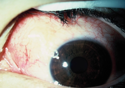
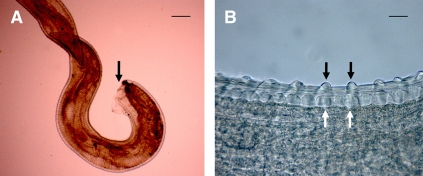
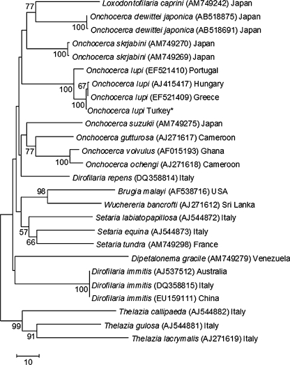
In the past decades, cases of canine ocular onchocercosis have been reported worldwide, particularly in the United States and Europe. Onchocerca lupi, originally described from a wolf, has been implicated in some of these cases, and its zoonotic role has been hypothesized on the basis of the reexamination of two cases of human ocular onchocerciasis. In the present study, we describe, for the first time, the occurrence of O. lupi in the subconjunctival region of the human eye in a patient from Turkey. The nematode was identified as O. lupi based on its morphology and molecular phylogenetic analysis of partial cox1 and 12S ribosomal DNA genes. The results suggest that O. lupi should be considered in the differential diagnosis of other eye parasitic infections in humans. The role of dogs as natural hosts of O. lupi and the vectors of this zoonotic parasite need to be investigated.
Figures
References
-
- Pisella PJ, Assaraf E, Rossaza C, Limon S, Baudouin C, Richard-Lenoble D. Conjunctivitis and ocular parasitic diseases. J Fr Ophtalmol. 1999;22:585–588. - PubMed
-
- Shen J, Gasser RB, Chu D, Wang Z, Yuan X, Cantacessi C, Otranto D. Human thelaziosis—a neglected parasitic disease of the eye. J Parasitol. 2006;92:872–875. - PubMed
-
- Anderson RC. Nematode Parasites of Vertebrates—Their Development and Transmission. New York: CAB International; 2000. pp. 417–422.
Publication types
MeSH terms
LinkOut - more resources
Full Text Sources
Medical
Miscellaneous
