

Malarial retinopathy in Bangladeshi adults
- PMID: 21212217
- PMCID: PMC3005494
- DOI: 10.4269/ajtmh.2011.10-0205
Malarial retinopathy in Bangladeshi adults
Abstract
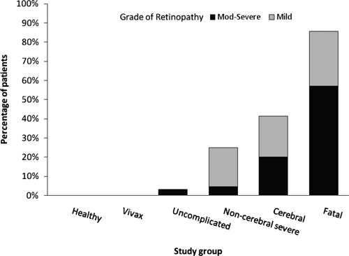
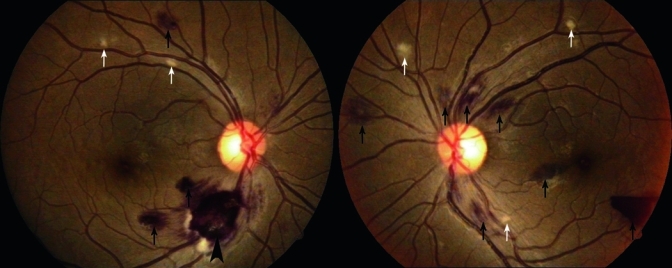
To establish if assessment of malarial retinopathy in adult malaria using ophthalmoscopy by non-ophthalmologists has clinical and prognostic significance, 210 Bangladeshi adults were assessed by both direct and indirect ophthalmoscopy; 20 of 20 healthy subjects and 20 of 20 patients with vivax malaria showed no retinal changes, whereas in patients with falciparum malaria, indirect ophthalmoscopy revealed malarial retinopathy (predominantly retinal hemorrhages) in 18 of 21 (86%) fatal, 31 of 75 (41%) cerebral, 16 of 64 (25%) non-cerebral but severe, and 1 of 31 (3%) uncomplicated cases. Direct ophthalmoscopy missed retinopathy in one of these cases and found fewer retinal hemorrhages (mean difference = 3.09; 95% confidence interval = 1.50-4.68; P < 0.0001). Severity of retinopathy increased with severity of disease (P for trend < 0.0001), and renal failure, acidosis, and moderate/severe retinopathy were independent predictors of mortality by both ophthalmoscopic techniques. Direct ophthalmoscopy by non-ophthalmologists is an important clinical tool to aid diagnosis and prognosis in adults with severe malaria, and indirect ophthalmoscopy by non-ophthalmologists, although more sensitive, provides minimal additional prognostic information.
Figures
References
-
- Lopez AD, Mathers CD, Ezzati M, Jamison DT, Murray CJ. Global and regional burden of disease and risk factors, 2001: systematic analysis of population health data. Lancet. 2006;367:1747–1757. - PubMed
-
- Poncet F. De la retino-choroidite palustre. Ann Ocul (Paris) 1878;79:201–218.
-
- Lewallen S, Taylor TE, Molyneux ME, Wills BA, Courtright P. Ocular fundus findings in Malawian children with cerebral malaria. Ophthalmology. 1993;100:857–861. - PubMed
Publication types
MeSH terms
Substances
Grants and funding
LinkOut - more resources
Full Text Sources
Medical
