. 2011 Mar 11;286(10):7865-7872.
doi: 10.1074/jbc.M110.191452. Epub 2011 Jan 6.

Kaposi sarcoma-associated herpesvirus degrades cellular Toll-interleukin-1 receptor domain-containing adaptor-inducing beta-interferon (TRIF)

Affiliations

Kaposi sarcoma-associated herpesvirus degrades cellular Toll-interleukin-1 receptor domain-containing adaptor-inducing beta-interferon (TRIF)

Humera Ahmad et al. J Biol Chem. .

Abstract

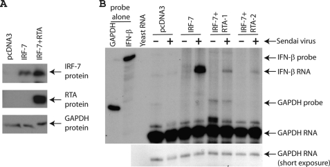
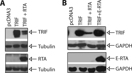
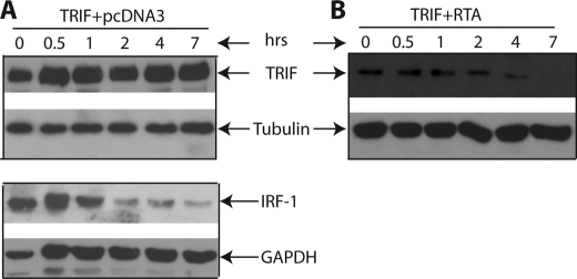
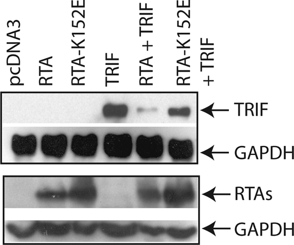
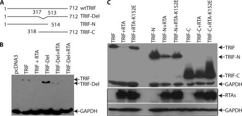
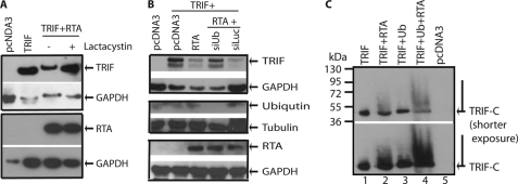
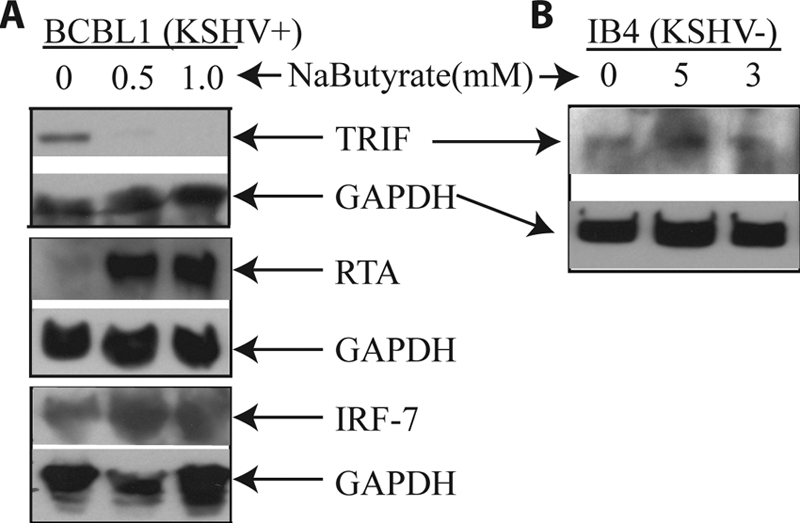
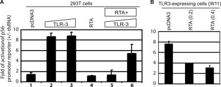
Kaposi sarcoma-associated herpesvirus (KSHV) is a human γ-herpesvirus associated with several human malignancies. The replication and transcription activator (RTA) is necessary and sufficient for the switch from KSHV latency to lytic replication. Toll-interleukin-1 receptor (TIR) domain-containing adaptor-inducing β-interferon (TRIF, also called TIR-domain-containing adaptor molecule-1 (TICAM-1)) is a signaling adaptor molecule that is critically involved in the Toll-like receptor 3 (TLR-3) and TLR-4 signaling pathways for type I interferon (IFN) production, a key component of innate immunity against microbial infection. In this report, we find a new mechanism by which RTA blocks innate immunity by targeting cellular TRIF. RTA specifically degrades TRIF by shortening the half-life of TRIF protein. This RTA-mediated degradation is at least partially mediated through the ubiquitin-proteasome pathway because proteasome inhibitors as well as knockdown of cellular ubiquitin expression alleviate the degradation. RTA may not directly interact with TRIF and may activate TRIF degradation indirectly through an unknown mediator(s). RTA targets multiple regions of TRIF and may use its ubiquitin ligase domain for the degradation. In addition, physiological levels of TRIF protein are down-regulated during KSHV lytic replication when RTA is expressed. Finally, RTA down-regulates double-stranded RNA-initiated activation of TLR-3 pathway, in the absence of degradation of IFN regulatory factor 7 (IRF-7). Taken together, these data suggest that KSHV employs a novel mechanism to block the innate immunity by degrading TRIF protein. This work may contribute to our understandings on how KSHV evades host immunity for its survival in vivo.


Figures

FIGURE 1.
FIGURE 1.
KSHV RTA blocks IFN production without IRF-7 degradation. A, RTA could not degrade IRF-7 in the 293T cells. IRF-7 expression plasmid (0.1 μg) plus various amounts of RTA (0.2 μg) were transfected into 293T cells in a 6-well plate. Total DNA for transfection was maintained the same with the use of vector DNA (pcDNA3). Lysates were used for Western blot analysis. The identity of proteins is as shown. B, RTA inhibits IFN production. IRF-7 expression plasmid (0.4 μg) plus RTA (0.8 μg) were transfected into 293T cells in a 10-cm dish. 1 day later, cells were infected with Sendai virus (20 HA units/ml) for 6 h. Total RNA was isolated, and RPA was performed with IFN-β and GAPDH probes. The identity of RNA is as shown.
FIGURE 2.
FIGURE 2.
RTA reduces the expression of TRIF protein. A, RTA reduces TRIF protein expression. 293T cells in 6-well plates were transfected with pcDNA3, RTA (0.2 μg), and TRIF expression plasmid (0.1 μg) in various combinations as shown on the top. Total DNA for transfection was maintained the same with the use of vector DNA. The cell lysates were obtained 1 day later for Western blot analysis. The identity of proteins is as shown. B, EBV RTA could not reduce the expression of TRIF. 293T cells were transfected with pcDNA3, RTA (0.2 μg), E-RTA (0.2 μg), and TRIF expression plasmid (0.1 μg) in various combinations as shown on the top. The cell lysates were obtained 1 day later. The membrane was stripped and probed with another antibody. The images in the same box indicate that they are derived from the same membranes. The identity of proteins is as shown.
FIGURE 3.
FIGURE 3.
RTA alters the TRIF protein stability. 293T cells in a 10-cm dish were transfected with TRIF (0.4 μg) (A) or TRIF plus RTA (0.8 μg) expression plasmid (B). Total DNA for transfection was maintained the same with the use of vector DNA. 6 h after transfection, cells were split into a 6-well plate. Cycloheximide (100 μg/ml) was added after a 12-h incubation. Cell lysates were made at various time intervals (in hours) as shown on the top, and Western blot analysis was performed. The membrane was stripped and probed with another antibody. The images in the same box indicate that they are derived from the same membranes. The identity of proteins is as shown.
FIGURE 4.
FIGURE 4.
An RTA mutant failed to reduce the protein expression of TRIF. 293T cells were transfected with vector pcDNA3, TRIF (0.1 μg), RTA (0.2 μg), and RTA-K152E (0.1 μg) expression plasmid in various combinations as shown on the top. Total DNA for transfection was maintained the same with the use of vector DNA. Cell lysates were made 1 day later, and Western blot analysis was performed. The membrane was stripped and probed with another antibody. The images in the same box indicate that they are derived from the same membranes. The identity of proteins is as shown.
FIGURE 5.
FIGURE 5.
RTA targets multiple regions of TRIF for degradation. A, schematic diagram of TRIF mutant constructs. The numbers denote the amino acid positions. The drawing is not to scale. B, TRIF-Del is degraded by RTA. 293T cells were transfected with vector pcDNA3, TRIF (0.1 μg), RTA (0.2 μg), and TRIF-Del mutant (0.1 μg) expression plasmids in various combinations as shown on the top. Total DNA for transfection was maintained the same with the use of vector DNA. Cell lysates were made 1 day later, and Western blot analysis was performed. Myc and GAPDH antibodies were used. The identity of proteins is as shown. C, both TRIF-N and TRIF-C are degraded by RTA. The two TRIF mutants, RTA, and RTA-K152E, were transfected into 293T cells with various combinations. FLAG, Myc, GAPDH, and RTA antibodies were used for the Western blot. The images in the same box indicate that they are derived from the same membranes. The identity of proteins is as shown.
FIGURE 6.
FIGURE 6.
RTA degrades TRIF through proteasome pathway. A, proteasome inhibitor alleviates TRIF degradation. 293T cells were transfected with pcDNA3, RTA (0.2 μg), and TRIF expression plasmid (0.1 μg) in various combinations as shown on the top. Total DNA for transfection was maintained the same with the use of vector DNA. 6 h after transfection, cells were washed and treated with lactacystin. The cell lysates were obtained 1 day later for Western blot analysis. The identity of proteins is as shown. B, knockdown of ubiquitin alleviates TRIF degradation. 293T cells were transfected with pcDNA3, RTA (0.1 μg), and TRIF (0.05 μg) expression plasmids in various combinations as shown on the top. In addition, siUb or siLuc (5 pmol of each) were also transfected. Cell lysates were made 1 day later, and Western blot analysis with TRIF antibody was performed. The membrane was stripped and probed with another antibody. The images in the same box indicate that they are derived from the same membranes. The identity of proteins is as shown. C, ubiquitin was added to TRIF-C. TRIF-C or RTA+TRIF-C were transfected with or without HA-Ub expression plasmid. The cell lysates were obtained 1 day later for Western blot analysis. More cell lysates were used for RTA+TRIF-C transfections. Two different exposures of the same membrane are shown. The identity of proteins is as shown.
FIGURE 7.
FIGURE 7.
TRIF is down-regulated during KSHV lytic replication. A, the inverse correlation between the expression of RTA and TRIF. BCBL1 cells were treated with sodium butyrate (NaButyrate) for 24 h. Cell lysates were made, and the expression of endogenous proteins was analyzed by Western blot analysis. The membrane was stripped and probed with another antibody. The same cell lysates were used, and images in the same box indicate that they are derived from the same membranes. The identity of proteins is as shown. B, sodium butyrate did not affect TRIF in KSHV-negative cell line (KSHV−). IB4 cells (KSHV−) were treated with sodium butyrate for 24 h, and the cell lysates were made. The expression of TRIF was examined.
FIGURE 8.
FIGURE 8.
RTA blocks TLR-3 signaling. A, RTA blocks TLR-3 signaling in 293T cells. Various plasmids were transfected into 293T cells as shown on the top. 0.1 or 0.2 μg of TLR-3 (columns 2, 3, 5, and 6) was used, respectively. RTA (0.2 μg) was used for transfection. Total DNA for transfection was maintained the same with the use of vector DNA. After 4–6 h of transfection, the cells were then treated (+) or not treated (−) with dsRNA (10 μg/ml). 1 day later, the cells were collected, and luciferase and β-gal assays were used for detection of reporter activation. The relative activation of p56 reporter (+dsRNA/−dsRNA) is as shown. Error bars indicate S.D. B, RTA blocks TLR-3 signaling in TLR-expressing cells. Various plasmids were transfected into TLR-3-expressing cells (WT11-9) as shown on the top. 0.2 or 0.4 μg of RTA was used, respectively. The cells were then treated (+) or not treated (−) with dsRNA (10 μg/ml). The relative activation of p56 reporter (+dsRNA/−dsRNA) is shown. One representative result is shown. Error bars indicate S.D.

References

    1. Moore P., Chang Y. (2001) in Virology (Knipe D. M. ed) pp. 2803–2833, Lippincott Williams and Wilkins, Philadelphia, PA
    1. Chang Y., Cesarman E., Pessin M. S., Lee F., Culpepper J., Knowles D. M., Moore P. S. (1994) Science 266, 1865–1869 - PubMed
    1. Chang Y., Moore P. S. (1996) Infect. Agents Dis. 5, 215–222 - PubMed
    1. West J. T., Wood C. (2003) Oncogene 22, 5150–5163 - PubMed
    1. Dourmishev L. A., Dourmishev A. L., Palmeri D., Schwartz R. A., Lukac D. M. (2003) Microbiol. Mol. Biol. Rev. 67, 175–212 - PMC - PubMed

Publication types

MeSH terms

LinkOut - more resources