. 2011 Feb 25;331(6020):1071-4.
doi: 10.1126/science.1199424. Epub 2011 Jan 6.

Global tissue revolutions in a morphogenetic movement controlling elongation

Affiliations

Global tissue revolutions in a morphogenetic movement controlling elongation

Saori L Haigo et al. Science. .

Abstract

Polarized cell behaviors drive axis elongation in animal embryos, but the mechanisms underlying elongation of many tissues remain unknown. Eggs of Drosophila undergo elongation from a sphere to an ellipsoid during oogenesis. We used live imaging of follicles (developing eggs) to elucidate the cellular basis of egg elongation. We find that elongating follicles undergo repeated rounds of circumferential rotation around their long axes. Follicle epithelia mutant for integrin or collagen IV fail to rotate and elongate, which results in round eggs. We present evidence that polarized rotation is required to build a polarized, fibrillar extracellular matrix (ECM) that constrains tissue shape. Thus, global tissue rotation is a morphogenetic behavior that uses planar polarity information in the ECM to control tissue elongation.


Figures

Figure 1
Figure 1. Follicle rotation coincides with elongation of the Drosophila egg
Anterior is left unless otherwise noted. Graphs report mean ± standard deviation. (A) Follicles elongate along the A-P axis during oogenesis. Collagen IV-GFP (green), F-actin (red), and DNA (cyan). (B) Follicle aspect ratio (AR) quantification reveals elongation primarily occurs between stages 5 and 9. Solid blue line indicates stages during which polarized rotation is observed. (C) Tracking of follicle cells (white and yellow dots, arrowheads) and germline nuclei (red; blue asterisks, arrows) rotate together. Note that the stage 9 follicle does not rotate. (D) Follicle cells (red; tracked cells outlined in blue) move against static Collagen IV-GFP fibrils (green, arrowheads). Scale bar in (A) 100 μm; (C) 50μm; (D) 10μm.
Figure 2
Figure 2. mys or vkg mosaic follicles do not undergo polarized rotation and do not elongate
(A-D) Follicles with epithelial clones of mys or vkg (GFP negative) show shape defects during rotation. mys mutant follicles (B) are rounder from stage 5; vkg mutant follicles (C) are rounder from stage 8. AR is displayed in (D). Asterisks indicate P<0.05. (E-G) Round follicles with mys (F) and vkg (G) clones are defective in rotation compared to control (E). WT cells (green intracellular); Indy-GFP (green membrane); tracked cells are pseudocolored red. (H) Percentage of follicles undergoing polarized, off-axis, or no rotation from live imaging. Scale bar in (A-C) 100 μm; (E-G) 25μm.
Figure 3
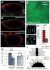
Figure 3. A polarized fibrillar Collagen IV matrix is built during follicle rotation
(A-F) Length and density of Collagen IV fibrils increase during follicle rotation. Collagen IV-GFP (green) puncta at stage 4 (A) mature into polarized fibrils (B-E). (F) Collagen fibrils orient perpendicular to the A-P axis. (G) Strategy to create RFP-marked follicle cells producing Collagen IV-GFP in genetic mosaics. Source cells distributed across the A-P axis (H) produce a uniform distribution of Collagen-GFP fibrils, while posteriorly-restricted source cells (I) produce only posterior labeled fibrils in stage 8 follicles. Scale bar in (A-E) 5 μm; (H-I) 25 μm.
Figure 4
Figure 4. A polarized fibrillar Collagen IV matrix maintains follicle shape
(A-C) Acute drug treatment of elongated stage 12 follicles. Collagenase (C) but not Latrunculin A (B) perturbs follicle shape, quantified in (D). (E-J) The Collagen IV matrix (green) is present but disorganized in round mys mutant follicles (compare E to Fig. 3C). Fibrils normally elongated in WT (F) fail to elongate in mys mutants (G); largest fibril shapes quantitated in (H). (I, J) Collagen fibril orientation is lost in mys mutant follicles. Double asterisks indicate P<0.01; triple asterisks indicate P≪0.001. Scale bar in (A-C) 100 μm; (E) 10 μm; (F-G) 3 μm.

References

    1. Keller R. Science. 2002;298:1950. - PubMed
    1. Andrew D, Ewald A. Dev Biol. 2010;341:34. - PMC - PubMed
    1. Wallingford J. Am J Med Genet C Semin Med Genet. 2005;135C:59. - PubMed
    1. Keller R. Development. 2006;133:2291. - PubMed
    1. Spradling A. In: The development of Drosophila melanogaster. Bate M, Martinez-Arias A, editors. CSHL Press; New York: 1993. pp. 1–70.

Publication types

MeSH terms

LinkOut - more resources