

Knee articular cartilage damage in osteoarthritis: analysis of MR image biomarker reproducibility in ACRIN-PA 4001 multicenter trial
- PMID: 21212364
- PMCID: PMC4104774
- DOI: 10.1148/radiol.10101174
Knee articular cartilage damage in osteoarthritis: analysis of MR image biomarker reproducibility in ACRIN-PA 4001 multicenter trial
Abstract
Purpose: To prospectively determine the reproducibility of quantitative magnetic resonance (MR) imaging biomarkers of the morphology and composition (spin lattice relaxation time in rotating frame [T1-ρ], T2) of knee cartilage in a multicenter multivendor trial involving patients with osteoarthritis (OA) and asymptomatic control subjects.
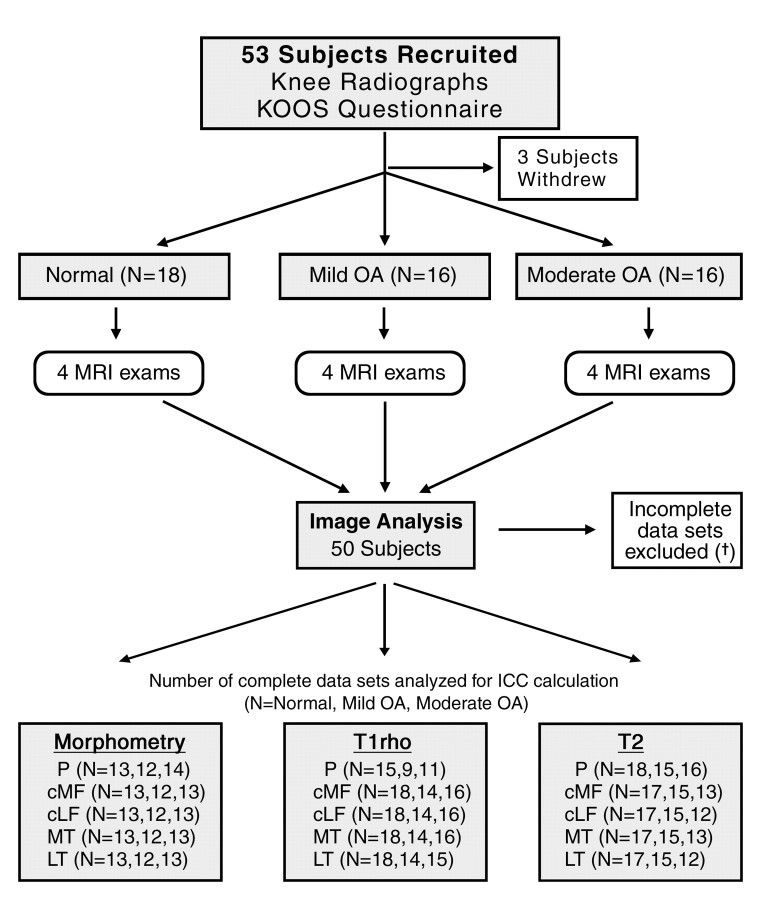
Materials and methods: This study was HIPAA compliant and approved by the institutional review committees of the participating sites, with written informed consent obtained from all participants. Fifty subjects from five sites who were deemed to have normal knee joints (n = 18), mild OA (n = 16), or moderate OA (n = 16) on the basis of Kellgren-Lawrence scores were enrolled. Each participant underwent four sequential 3-T knee MR imaging examinations with use of the same imager and with 2-63 days (median, 18 days) separating the first and last examinations. Water-excited three-dimensional T1-weighted gradient-echo imaging, T1-ρ imaging, and T2 mapping of cartilage in the axial and coronal planes were performed. Biomarker reproducibility was determined by using intraclass correlation coefficients (ICCs) and root-mean-square coefficients of variation (RMS CVs, expressed as percentages).
Results: Morphometric biomarkers had high reproducibility, with ICCs of 0.989 or greater and RMS CVs lower than 4%. The largest differences between the healthy subjects and the patients with radiographically detected knee OA were those in T1-ρ values, but precision errors were relatively large. Reproducibility of T1-ρ values was higher in the thicker patellar cartilage (ICC range, 0.86-0.93; RMS CV range, 14%-18%) than in the femorotibial joints (ICC range, 0.20-0.84; RMS CV range, 7%-19%). Good to high reproducibility of T2 was observed, with ICCs ranging from 0.61 to 0.98 and RMS CVs ranging from 4% to 14%.
Conclusion: MR imaging measurements of cartilage morphology, T2, and patellar T1-ρ demonstrated moderate to excellent reproducibility in a clinical trial network.
© RSNA, 2011.
Figures
References
-
- Eckstein F, Mosher T, Hunter D. Imaging of knee osteoarthritis: data beyond the beauty. Curr Opin Rheumatol 2007;19(5):435–443. - PubMed
-
- Le Graverand MP, Buck RJ, Wyman BT, et al. . Change in regional cartilage morphology and joint space width in osteoarthritis participants versus healthy controls: a multicentre study using 3.0 Tesla MRI and Lyon-Schuss radiography. Ann Rheum Dis 2010;69(1):155–162. - PubMed
-
- Abadie E, Ethgen D, Avouac B, et al. . Recommendations for the use of new methods to assess the efficacy of disease-modifying drugs in the treatment of osteoarthritis. Osteoarthritis Cartilage 2004;12(4):263–268. - PubMed
-
- Eckstein F, Buck RJ, Burstein D, et al. . Precision of 3.0 Tesla quantitative magnetic resonance imaging of cartilage morphology in a multicentre clinical trial. Ann Rheum Dis 2008;67(12):1683–1688. - PubMed
Publication types
MeSH terms
Substances
Grants and funding
LinkOut - more resources
Full Text Sources
Other Literature Sources
Medical
