Review
. 2011 Jan 7;108(1):98-112.
doi: 10.1161/CIRCRESAHA.110.223586.

Alternans and arrhythmias: from cell to heart

Affiliations
Review

Alternans and arrhythmias: from cell to heart

James N Weiss et al. Circ Res. .

Abstract

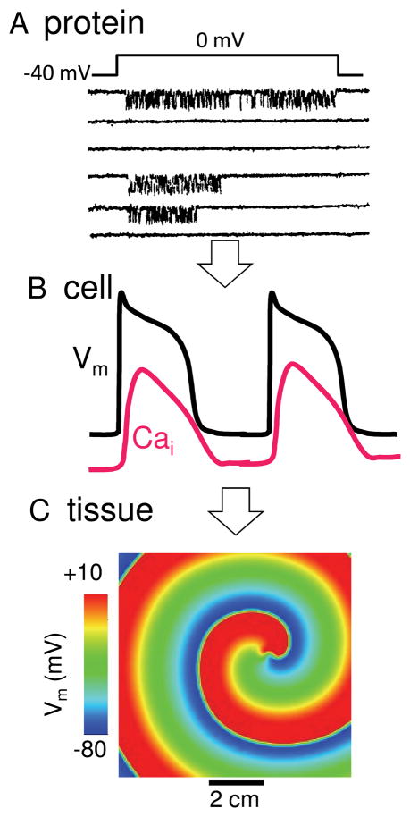
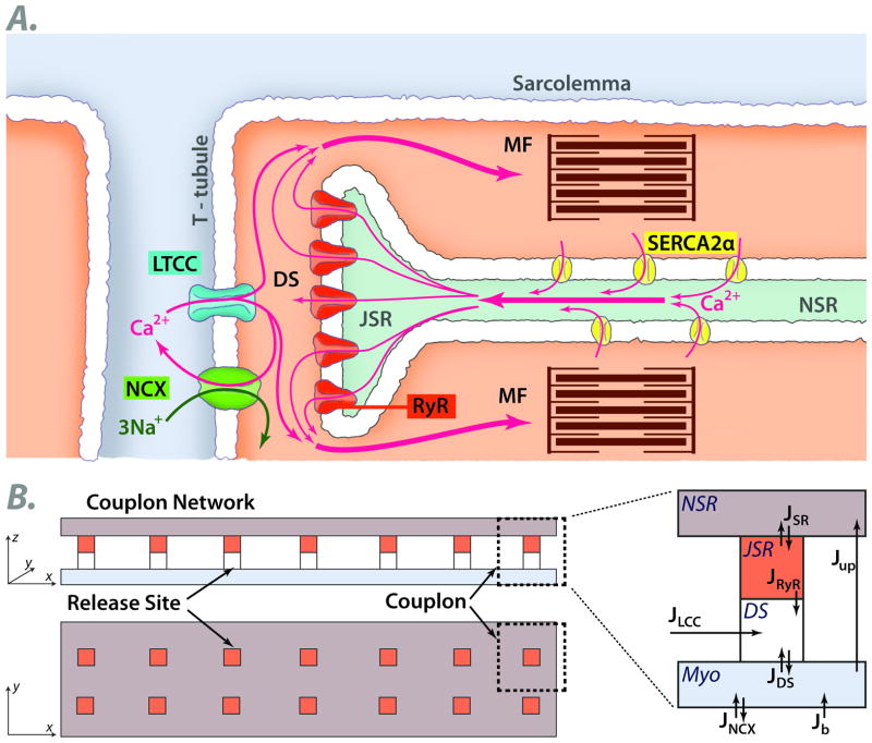
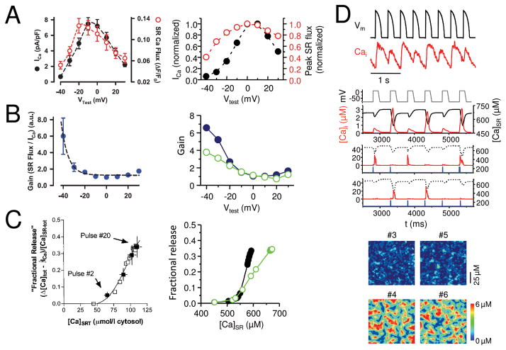
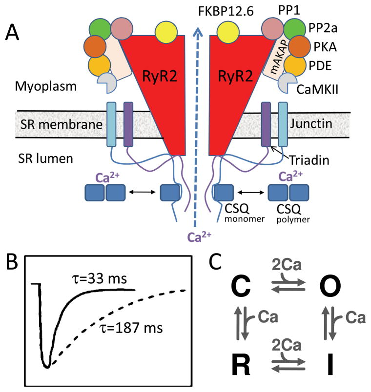
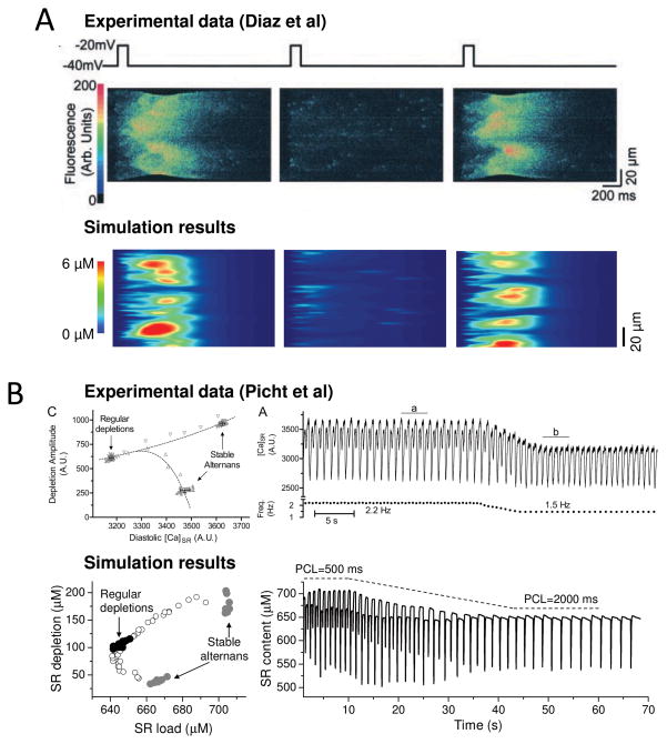
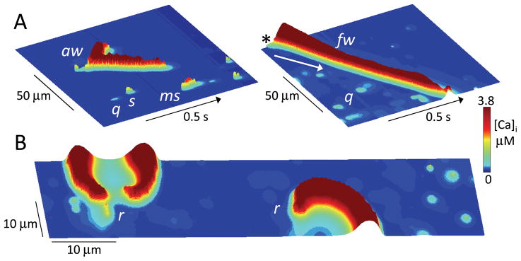
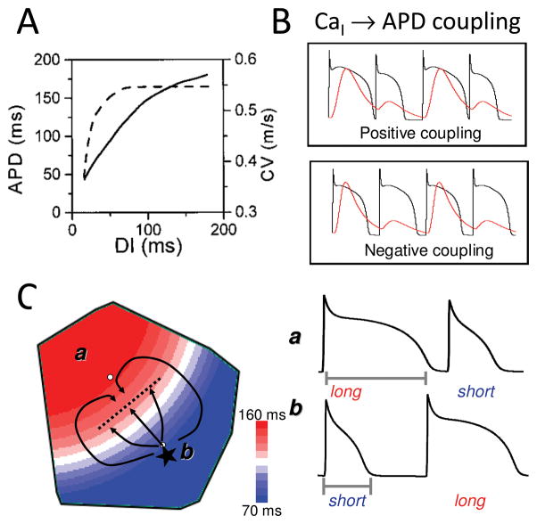
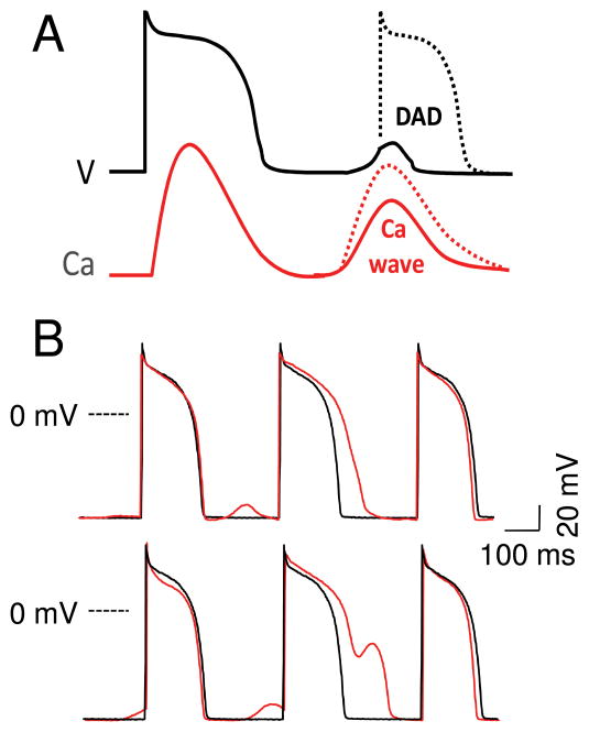
The goal of systems biology is to relate events at the molecular level to more integrated scales from organelle to cell, tissue, and living organism. Here, we review how normal and abnormal excitation-contraction coupling properties emerge from the protein scale, where behaviors are dominated by randomness, to the cell and tissue scales, where heart has to beat with reliable regularity for a lifetime. Beginning with the fundamental unit of excitation-contraction coupling, the couplon where L-type Ca channels in the sarcolemmal membrane adjoin ryanodine receptors in the sarcoplasmic reticulum membrane, we show how a network of couplons with 3 basic properties (random activation, refractoriness, and recruitment) produces the classic physiological properties of excitation-contraction coupling and, under pathophysiological conditions, leads to Ca alternans and Ca waves. Moving to the tissue scale, we discuss how cellular Ca alternans and Ca waves promote both reentrant and focal arrhythmias in the heart. Throughout, we emphasize the qualitatively novel properties that emerge at each new scale of integration.


Figures

Fig. 1
Fig. 1
A. Stochastic protein behavior. Single channel recording of an L-type Ca channel from a cardiac myocyte, during successive voltage clamp pulses from −40 to 0 mV, showing different behaviors on each sweep. Downward deflections indicate channel openings. B. Regular cell behavior. Integrated behavior of stochastic ion channel network creates a dependably regular AP and Ca transient from beat-to-beat. C. Pathological tissue behavior. In addition to coordinating the normal heart beat, wave propagation at the tissue level also permits reentry which can cause life-threatening arrhythmias.
Fig. 2
Fig. 2
A. Schematic of a cardiac couplon, formed by L-type Ca channels (LTCC) in the T-tubular membrane and ryanodine receptors (RyR) in the apposed junctional SR (JSR). Extracellular Ca entering through LTCC triggers RyR to open, releasing SR Ca into the myoplasm (MYO) to activate the myofilaments (MF). Ca is then pumped back into the nonjuctional SR (NSR) by a Ca pump (SERCA2a) or extruded from the cell via Na-Ca exchange (NCX). B. Spatially-distributed 2D model of the couplon network. See text. DS=dyadic space. J=Ca fluxes between compartments.
Fig. 3
Fig. 3
A. Graded Ca release. SR Ca release flux (black) tracks the amplitude of the L-type Ca current (red) during voltage clamps to different membrane voltages (Vm) in a rabbit ventricular myocyte , reproduced by the couplon network model(right). B. Voltage-dependent EC coupling gain. EC coupling gain, defined as the ratio of SR Ca release to the L-type Ca current amplitude, is higher at less depolarized voltages in experimental data from rabbit ventricular myocytes (left, from 25). The steep decline in gain is reproduced better in the couplon network model(right) when the couplons are coupled (solid symbols) than when uncoupled (open symbols). C. Steep SR fractional release-load relationship. The fraction of SR Ca released increases steeply as the SR load increases in a rabbit ventricular myocyte (left). The steepness is more accurately reproduced by the couplon network model(right) when the couplons are coupled (solid symbols) than when uncoupled (open symbols). D. Ca alternans. During rapid pacing with a fixed AP wavefrom (black), the Ca transient (red) alternates between large and small on successive beats in a patch-clamped rabbit ventricular myocyte (top panel), reproduced by the couplon network model(lower panels). 2nd panel shows alternans of the global Ca transient and SR Ca content during pacing with a fixed voltage waveform. 3rd and 4th panels show two representative couplons in the network, exhibiting irregular activations instead of alternans. 5th and 6th panels show the spatial patterns of Ca release from couplons during alternans on 4 successive beats. Note that when the two small beats or two large beats are compared to each other, the spatial patterns differ, indicating the macroscopic alternans is not accompanied by microscopic alternans.
Fig. 4
Fig. 4
A. Ryanodine receptor macromolecular complex. The cardiac ryanodine receptor (RyR2) is a tetramer which forms a macromolecular complex with multiple interacting partners, including anchoring proteins (mAKAP + others not shown), protein kinases and phosphatases (PKA, CaMKII, PP1, PP2a), and other additional accessory regulatory proteins (FKBP12.6, triadin, junction, calsequestran, CSQ). B. Couplon refractoriness. Time course of SR refilling (solid line) vs couplon recovery from inactivation (dashed line), from Brochet et al . C. Simple four-state RyR model, from Stern , without SR luminal Ca regulation.
Fig. 5
Fig. 5
A. Ca alternans due to CICR waves on alternate beats, elicited by successive voltage clamp pulses from −40 to −20 mV in a ventricular myocyte (above, from 6) and the couplon network model(below). Panels show line scans, with spatial position vertically and time horizontally. Note that the Ca waves initiate at different locations on the 1st and 3rd beats. B. Dissociation between SR Ca release and load during alternans. When heart rate was decreased (left panels) , Ca alternans resolved in a patch-clamped rabbit ventricular myocyte (above, from 32) and the 3 R model (below). Left panels show that SR load (diastolic [Ca]SR) was lower during regular beating than during alternans, even though the SR depletion was larger than during the small Ca transient.
Fig. 6
Fig. 6. Ca signaling hierarchy in the couplon network
A. Ca quarks (q), sparks (s), macrosparks (ms), aborted wave (aw) and full wave (fw), shown as labeled in line scans of cytoplasmic [Ca] along a line through the center of the couplon network array (100 x 20 μm) versus time. Cytoplasmic free [Ca] is indicated by height and color scale. In the right panel, the SR Ca load was higher to promote the full wave, which started as a spark in the upper corner (asterix) and propagated downward (arrow) by CICR through the full length of the couplon array. B. Snapshot of Ca rotors (r) in a couplon array (100 x 20 μm) at high SR Ca load. Figure-eight spiral wave reentry causing a double rotor is shown at left, with a single spiral wave rotor at right.
Fig. 7
Fig. 7
A. APD and CV restitution curves. As diastolic interval (DI) decreases, APD shortens and CV slows. B. Positive and negative Cai-APD coupling. See text. C. Arrhythmogenic spatially discordant APD alternans. In region A, APD alternans has a long-short pattern, whereas region B has a short-long pattern, separated by a nodal line without alternans. If a PVC (*) occurs in the short APD region, it can block as it propagates across the nodal line into in the long APD region, while propagating laterally until the long APD region recovers, initiating figure-eight reentry.
Fig. 8
Fig. 8
A. Sub- and supra-threshold DADs. Depending on the size and rate of rise of the Ca transient during the Ca wave(s), and the diastolic Ca-voltage coupling gain, a DAD can remain below or above the threshold to trigger an AP. B. DAD-repolarization interaction. DADs recorded from a paced isolated rabbit ventricular myocyte before (black traces) and after (red traces) exposure to isoproterenol. Depending on the timing, subthreshold DADs can cause APD prolongation (upper) and frank EADs (lower).

References

    1. Stern MD. Theory of excitation-contraction coupling in cardiac muscle. Biophys J. 1992;63(2):497–517. - PMC - PubMed
    1. Cheng H, Lederer WJ, Cannell MB. Calcium sparks: elementary events underlying excitation-contraction coupling in heart muscle. Science. 1993;262(5134):740–744. - PubMed
    1. Lipp P, Niggli E. A hierarchical concept of cellular and subcellular Ca(2+)-signalling. Progress in Biophysics and Molecular Biology. 1996;65(3):265–296. - PubMed
    1. Wier WG, Egan TM, Lopez-Lopez JR, Balke CW. Local control of excitation-contraction coupling in rat heart cells. J Physiol. 1994;474(3):463–471. - PMC - PubMed
    1. Eisner DA, Diaz ME, Li Y, O'Neill SC, Trafford AW. Stability and instability of regulation of intracellular calcium. Exp Physiol. 2005;90(1):3–12. - PubMed

Publication types

Substances