Case Reports
. 2011;8(4):221-9.
doi: 10.1159/000322228. Epub 2011 Jan 5.

Presence of reactive microglia and neuroinflammatory mediators in a case of frontotemporal dementia with P301S mutation

Affiliations
Case Reports

Presence of reactive microglia and neuroinflammatory mediators in a case of frontotemporal dementia with P301S mutation

Arianna Bellucci et al. Neurodegener Dis. 2011.

Abstract

Background: Recent findings, showing the presence of an inflammatory process in the brain of transgenic mice expressing P301S mutated human tau protein, indicate that neuroinflammation may contribute to tau-related degeneration in frontotemporal dementia and parkinsonism linked to chromosome 17 with tau mutations (FTDP-17T).

Objective: To investigate the occurrence of neuroinflammatory changes in the brain of a patient affected by FTDP-17T associated with the P301S mutation and showing a frontotemporal dementia phenotype as well as in the brain of a patient affected by another FTDP-17T phenotype: multiple system tauopathy with presenile dementia.

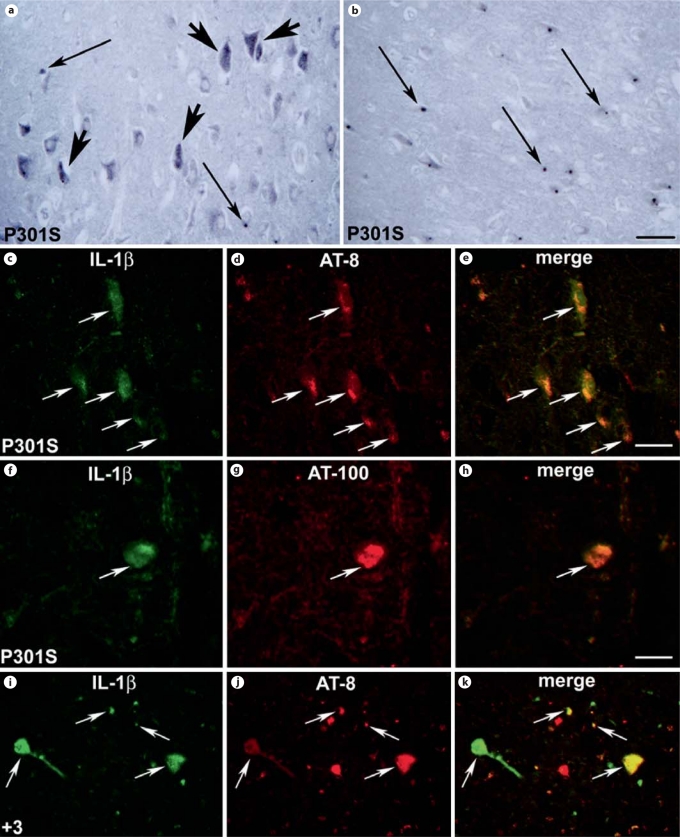
Methods: We used immunohistochemical methods to visualize activated microglia, interleukin-1b (IL-1b)-, cyclooxygenase-2 (COX-2)-expressing cells.

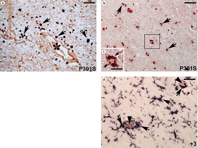
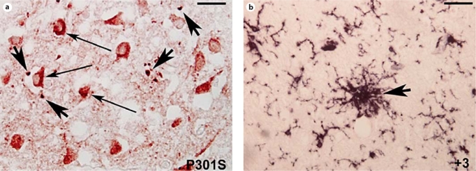
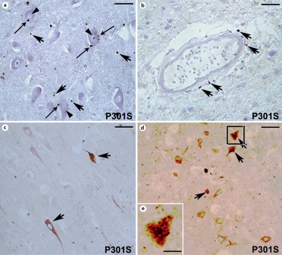
Results: In the brain of the patient with the P301S mutation, a strong neuroinflammatory reaction was present. Activated microglia/infiltrating macrophages expressing the cluster of differentiation 68 and major histocampatibility complex class II cell surface receptors, encoded by the human leukocyte antigen DP-DQ-DR, were detected in the cortex and hippocampus. IL-1b and COX-2 expression were induced in neuronal and glial cells. These neuroinflammatory changes were different from those observed in the brain of the patient bearing the +3 mutation, where macrophage infiltration was absent, microglial cells displayed an earlier stage of activation and COX-2 was not detected.

Conclusions: Our findings suggest that microglial activation and the production of proinflammatory mediators by phospho-tau-positive neurons and glial cells may differentially contribute to neuronal death and disease progression in neurodegenerative tauopathies.


Figures

Fig. 1
Fig. 1
CD68 (black staining) and AP-422 (red staining; colors in the online version only) double labeling. a CD68-positive roundshaped cells (arrows) in the hippocampal white matter of the patient with the P301S mutation. Note the abundance of these cells in the surroundings of the blood vessel (asterisk) indicative of macrophage infiltration. b CD68-immunoreactive cells (arrows) cluster around AP-422-positive neurons (red-labeled cells) in the hippocampal gray matter of the P301S patient. c High magnification of the square in b showing the round-shaped morphology of CD68-positive cells (arrows) in the surroundings of an AP-422-positive neuron. d CD68 (black staining) and AP-422 (red staining) double labeling. In the brain of the patient with the +3 mutation, CD68-antibody-labeled cells with swollen cell bodies and thick shortened but still ramified processes in the parenchyma and in the surroundings of AP-422-positive neurons (indicated by the arrowheads). a, b, d Scale bar = 100 μm; c scale bar = 40 μm.
Fig. 2
Fig. 2
HLA-DP-DQ-DR (black staining) and AP-422 (red staining; colors in the online version only) double labeling. a Cortex of the patient with the P301S mutation. Note the presence of numerous cells with ameboid morphology (large arrows) in the surroundings of AP-422-positive neurons (thin arrows, red cells). b Cortex of the patient affected by FTDP-17T with the +3 mutation. Note the presence of clusters of bushy activated HLA-DPDQ-DR-positive microglial cells (arrow). a Scale bar = 50 μm; b scale bar = 80 μm.
Fig. 3
Fig. 3
IL-1β-positive cells in the brain of the patients with P301S (a–h) and +3 (i–k) mutations. a IL-1β-staining in neuronal (large arrows) cells in the cortical gray matter. Please note the presence of numerous IL-1β-positive dots (thin arrows). b IL-1β-positive round-shaped dots in the white matter of the cortex (thin arrows). c, d Double-labeling immunofluorescence staining for IL-1β (green; colors in the online version only) and tau phosphorylated at S202 and T205 (antibody AT-8; red) in the hippocampus of the patient with the P301S mutation. e Merged image of the pictures in c and d showing the expression of IL-1β within the AT-8-positive neurons that is confirmed by the yellow color. f, g Doublelabeling immunofluorescence staining for IL-1β (green) and tau phosphorylated at T212 and S214 (antibody AT-100; red) in the hippocampus of the patient with the P301S mutation. h Merged image of the pictures in f and g showing the expression of IL-1β within the AT-100-positive neurons that is confirmed by the yellow color. i, j Double-labeling immunofluorescence staining for IL-1β (green) and AT-8-positive tau protein in the cortex of the patient with the +3 mutation. k Merged image of the pictures in i and j showing expression of IL-1β within some of the AT-8-positive neurons and degenerating neuritis as confirmed by the yellow color (arrows). a, i–k Scale bar = 150 μm; b scale bar = 210 μm; c–e scale bar = 100 μm; f–h scale bar = 80 μm.
Fig. 4
Fig. 4
COX-2 immunoreactivity in the brain of the patient with the P301S mutation. a COX-2-immunopositive round-shaped macrophage-like cells in the parenchyma (large arrows) and around (thin arrows) neurons (arrowheads) in the cortex. b Note the presence of COX-2-positive macrophage-like cells in the surroundings of blood vessels (arrows). c Some COX-2-positive neurons are present in the gray matter of the hippocampus (arrows). d Double labeling for COX-2 (black) and tau phosphorylated at S202 and T205 (antibody AT-8; red in the online version only) in the hippocampus. Note the presence of COX-2 staining in phosphorylated-tau-positive neurons (arrow). e Higher magnification of the square in d. Please note the presence of the COX-2 dark labeling in the phosphorylated-tau-po sitive neuron. a Scale bar = 80 μm; b scale bar = 150 μm; c scale bar = 165 μm; d scale bar = 170 μm. e scale bar = 35 μm.

References

    1. Wszolek ZK, Tsuboi Y, Farrer M, Uitti RJ, Hutton ML. Hereditary tauopathies and parkinsonism. Adv Neurol. 2003;91:153–163. - PubMed
    1. Baba Y, Baker MC, Le Ber I, Brice A, Maeck L, Kohlhase J, Yasuda M, Stoppe G, Bugiani O, Sperfeld AD, Tsuboi Y, Uitti RJ, Farrer MJ, Ghetti B, Hutton ML, Wszolek ZK. Clinical and genetic features of families with frontotemporal dementia and parkinsonism linked to chromosome 17 with a P301S tau mutation. J Neural Transm. 2007;114:947–950. - PubMed
    1. Gasparini L, Terni B, Spillantini MG. Frontotemporal dementia with tau pathology. Neurodegener Dis. 2007;4:236–253. - PubMed
    1. Bugiani O, Murrell JR, Giaccone G, Hasegawa M, Ghigo G, Tabaton M, Morbin M, Primavera A, Carella F, Solaro C, Grisoli M, Savoiardo M, Spillantini MG, Tagliavini F, Goedert M, Ghetti B. Frontotemporal dementia and corticobasal degeneration in a family with a P301S mutation in tau. J Neuropathol Exp Neurol. 1999;58:667–677. - PubMed
    1. Bugiani O. FTDP-17: phenotypical heterogeneity within P301S. Ann Neurol. 2000;48:126. - PubMed

Publication types