Review
. 2011 Feb;58(2):191-205.
doi: 10.1007/s12630-010-9419-9. Epub 2011 Jan 7.

Anesthetic sites and allosteric mechanisms of action on Cys-loop ligand-gated ion channels

Affiliations
Review

Anesthetic sites and allosteric mechanisms of action on Cys-loop ligand-gated ion channels

Stuart A Forman et al. Can J Anaesth. 2011 Feb.

Abstract

Purpose: The Cys-loop ligand-gated ion channel superfamily is a major group of neurotransmitter-activated receptors in the central and peripheral nervous system. The superfamily includes inhibitory receptors stimulated by γ-aminobutyric acid (GABA) and glycine and excitatory receptors stimulated by acetylcholine and serotonin. The first part of this review presents current evidence on the location of the anesthetic binding sites on these channels and the mechanism by which binding to these sites alters their function. The second part of the review addresses the basis for this selectivity, and the third part describes the predictive power of a quantitative allosteric model showing the actions of etomidate on γ-aminobutyric acid type A receptors (GABA(A)Rs).

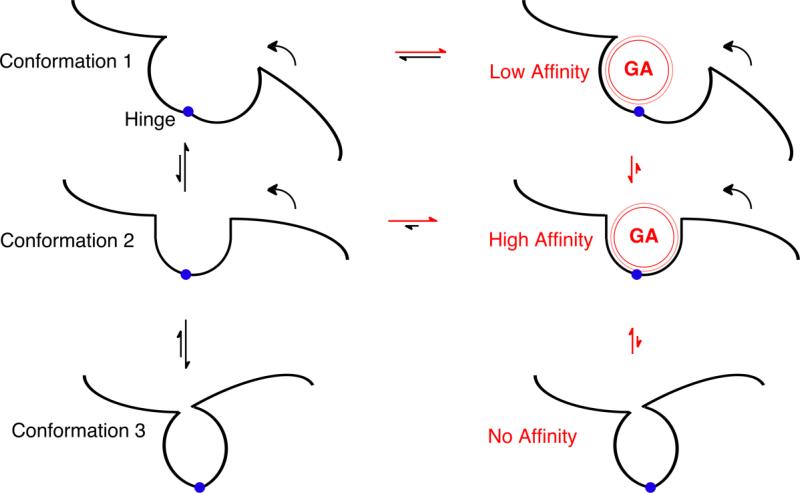
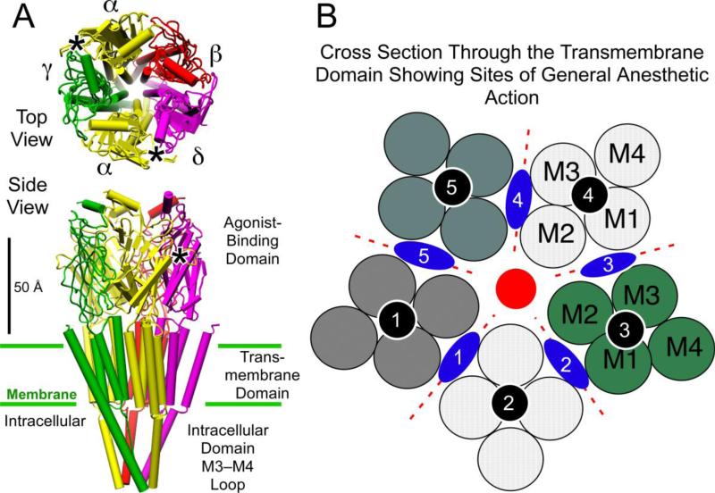
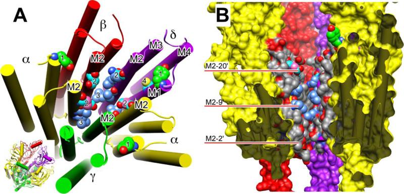
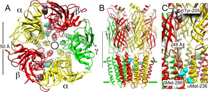
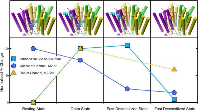
Principal findings: General anesthetics at clinical concentrations inhibit the excitatory receptors and enhance the inhibitory receptors. The location of general anesthetic binding sites on these receptors is being defined by photoactivable analogues of general anesthetics. The receptor studied most extensively is the muscle-type nicotinic acetylcholine receptor (nAChR), and progress is now being made with GABA(A)Rs. There are three categories of sites that are all in the transmembrane domain: 1) within a single subunit's four-helix bundle (intrasubunit site; halothane and etomidate on the δ subunit of AChRs); 2) between five subunits in the transmembrane conduction pore (channel lumen sites; etomidate and alcohols on nAChR); and 3) between two subunits (subunit interface sites; etomidate between the α1 and β2/3 subunits of the GABA(A)R).

Conclusions: These binding sites function allosterically. Certain conformations of a receptor bind the anesthetic with greater affinity than others. Time-resolved photolabelling of some sites occurs within milliseconds of channel opening on the nAChR but not before. In GABA(A)Rs, electrophysiological data fit an allosteric model in which etomidate binds to and stabilizes the open state, increasing both the fraction of open channels and their lifetime. As predicted by the model, the channel-stabilizing action of etomidate is so strong that higher concentrations open the channel in the absence of agonist. The formal functional paradigm presented for etomidate may apply to other potent general anesthetic drugs. Combining photolabelling with structure-function mutational studies in the context of allosteric mechanisms should lead us to a more detailed understanding of how and where these important drugs act.


Figures

Figure 1
Figure 1. Principles of Allosterism
The black lines depict part of a protein in a region where two domains are joined by a structural hinge (blue circle). In the left hand column, the protein can adopt three different conformations depending on movement of the right hand domain around the hinge (curved arrows). The relative size of the two black vertical straight arrows between each pair of conformations suggest the equilibrium distribution between the conformations; Conformations 1 and 3 are favored relative to Conformation 2. In the right hand column, general anesthetics (red sphere, GA) have the opportunity to bind to each of these conformations in the pocket between the domains. Because the intermolecular dispersion forces between the general anesthetic and the protein are very short range (depicted by the thin red outer line), strong, high affinity interactions only occur when the anesthetic fits snugly in the pocket (i.e. in Conformation 2). If the hinge closes too far (Conformation 3), steric hindrance prevents the anesthetic from binding. The horizontal red and black arrows depict how anesthetic binding perturbs the equilibrium between anesthetic free (left) and anesthetic bound (right) protein. In this example, Conformation 2 is sufficiently stabilized by the anesthetic's binding energy, that it is now the most stable relative to the Conformation 1 and 3.
Figure 2
Figure 2
General anesthetic binding sites on ligand-gated ion channels of the Cys-loop receptor superfamily. Panel A shows the structure of the nicotinic acetylcholine receptor (Unwin 2005), with its five subunits arranged centro-symmetrically around a central ion pore or channel, in both top view from the extracellular side and side view. The agonist site (*)in the extracellular agonist-binding domain is on the a-subunits in the interface with the g- and d-subunits. Panel B shows a schematic of a cross section through the transmembrane region. Each subunit, separated by dotted red lines, consists of four transmembrane helices shown as circles and numbered in the order they appear in the sequence. Three categories of anesthetic binding site (see text) are superimposed; intrasubunit sites (black circles); a channel lumen site (red circle), and subunit interface sites (blue lozenges). Molecular graphics images were produced using the UCSF Chimera package from the Resource for Biocomputing, Visualization, and Informatics at the University of California, San Francisco (supported by NIH P41 RR-01081) (74).
Figure 3
Figure 3
Formulae of photolabels referred to herein.
Figure 4
Figure 4
Residues photolabeled on nicotinic receptors by three classes of general anesthetics. Panel A shows a slightly tilted top view of just the transmembrane domain of the nAChR of the nicotinic acetylcholine receptor (nAChR) from Torpedo using the cryoelectron microscopy structure (20). The agonist-binding domain has been omitted for clarity, but is shown in the inset at bottom right, where the orientation is the same as in the larger diagram. The subunits are color-coded and labeled as in Fig. 2. The helices are shown as rods and the atoms of photolabeled residues, including the backbone atoms, are shown in space-filled mode. All other residues are omitted for clarity. Oxygen (red) and nitrogen (blue) atoms are colored conventionally. The carbon atoms are color coded to denote which anesthetics photolabeled the residue: green, halothane; cyan, azietomidate; cornflower blue, TDBzl-etomidate; salmon, azioctanol. In some cases, more than one agent photolabels the same residue and individual carbons are given different colors accordingly. Number code: 1. αY213 & δY228 photolabeled on M1 by halothane; 2. αE262, βD268 & δQ276 (the M2-20' residues) photolabeled by azietomidate and azioctanol; 3. αL251, δL265 (the M2-9' residues) photolabeled by TDBzl-etomidate; 4. δC236 on M1 Photolabeled by azietomidate. Panel B shows a cross-section through the transmembrane domain of the same nAChR structure. The γ-subunit has been removed to facilitate a view of the ion pore. The viewer is situated at the γ-subunit and the two α-subunits are closest to the viewer. The subunits have the same color code as before but are presented surfaced. The dark grey regions denote where the surface of the α-subunits have been cut through and serve to emphasize the free space that exists within the 4-helix bundle of subunits. The central ion channel is open to view. The M2 helices are colored conventionally with grey for carbon, red for oxygen and blue for nitrogen, except that in photolabeled residues some of the carbons have the same color code as in panel A. The red bars point to residues on M2 using the prime numbering system, where 1' is the residue following the last charged residue before the M2 helix and M2-9' is always the conserved leucine.
Figure 5
Figure 5
Azietomidate photolabels the GABAAR in the transmembrane domain between the a- and b-subunits. Panel A shows a top view from the extracellular side of the α1β2γ2L GABAAR homology model of Li et al (25). The five subunits, which are consistently color coded in all panels, are arranged centrosymmetrically around the pore, denoted by a circle. Panel B shows the same receptor in side view; the M3–M4 intracellular loop is omitted in this model. The scale bar is 50 Å in both pannels. Panel C shows a detail of Panel B. The partly obscured scale bar is 20 Å. Only the secondary structure is shown except for two sets of residues. The residues with carbons shown in cyan are those photolabeled by azietomidate, αMet-236 and βMet-286, and that with dark grey carbons is βTyr-205, which is a residue in the GABA–binding pocket, nearly 50 Å away.
Figure 6
Figure 6
The degree of photolabeling of three sites on the nAChR varies with the receptor's conformation, supporting allosteric action. The graph shows the relative level of photoincorporation for three different sites on the nicotinic acetylcholine receptor. The photoincorporation level is normalized to that first observed (either the resting or the open state). The upper panel depicts the transmembrane domain of the nAChR and the photolabeled residues in each state. The subunit colors are yellow for α, green for γ, and purple for δ. The β-subunit is shown in outline only to allow the channel residues to be seen. The carbon atoms are colored with the same color as the symbols on the graph. At two sites photolabeling is negligible in the resting state but increases dramatically when the channel opens. One of these sites (orange triangles) is in the upper part of the channel (M2–20') and the other (cyan squares) is in the intrahelical bundle of the β-subunit. The behavior of these sites diverges during desensitization. The channel site changes modestly, whereas the intrahelical site remains unchanged during fast desensitization and then decreases dramatically upon slow desensitization. The third site (blue circles) is also in the channel but at the conserved M2–9' leucines. It behaves differently from the site in the upper part of the channel. It is photolabeled in the resting state and in the open state, but photolabeling decreases dramatically during fast desensitization and remains unchanged during slow desensitization. Data for azietomidate (triangles) is from (40) and for TID (circles and squares) is from (52, 53).
Figure 7
Figure 7
The scheme depicts equilibrium two-state allosteric co-agonism for GABA and etomidate actions on GABAA receptors, as described by Rüsch et al (15). There are two equivalent GABA sites and two equivalent etomidate sites. Only doubly-bound states are shown, both for simplicity and because they are the most highly populated states when ligands are present. GABA binding transitions are blue, etomidate binding transitions are red, and gating (opening and closing) transitions are black. The L0 parameter describes the basal equilibrium between closed (R) and open (O) states. KG is the dissociation constant for GABA interactions with R-state receptors and KG* is the dissociation constant for GABA interactions with O-state receptors. The GABA efficacy factor, c, is defined as KG*/KG. KE is the dissociation constant for etomidate interactions with R-state receptors and KE* is the dissociation constant for etomidate interactions with O-state receptors. The anesthetic efficacy factor, d, is defined as KE*/KE.

References

    1. Jurd R, Arras M, Lambert S, et al. General anesthetic actions in vivo strongly attenuated by a point mutation in the GABA(A) receptor beta3 subunit. FASEB Journal. 2003;17:250–2. - PubMed
    1. Campagna JA, Miller KW, Forman SA. Mechanisms of actions of inhaled anesthetics. N Engl J Med. 2003;348:2110–24. - PubMed
    1. Hemmings HC, Jr., Akabas MH, Goldstein PA, Trudell JR, Orser BA, Harrison NL. Emerging molecular mechanisms of general anesthetic action. Trends Pharmacol Sci. 2005;26:503–10. - PubMed
    1. Rudolph U, Antkowiak B. Molecular and neuronal substrates for general anaesthetics. Nat Rev Neurosci. 2004;5:709–20. - PubMed
    1. Grasshoff C, Drexler B, Rudolph U, Antkowiak B. Anaesthetic drugs: linking molecular actions to clinical effects. Curr Pharm Des. 2006;12:3665–79. - PubMed

Publication types

MeSH terms