Sequential dissection of multiple ionic currents in single cardiac myocytes under action potential-clamp
- PMID: 21215755
- PMCID: PMC3047417
- DOI: 10.1016/j.yjmcc.2010.12.020
Sequential dissection of multiple ionic currents in single cardiac myocytes under action potential-clamp
Abstract
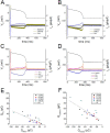
The cardiac action potential (AP) is shaped by myriad ionic currents. In this study, we develop an innovative AP-clamp Sequential Dissection technique to enable the recording of multiple ionic currents in the single cell under AP-clamp. This new technique presents a significant step beyond the traditional way of recording only one current in any one cell. The ability to measure many currents in a single cell has revealed two hitherto unknown characteristics of the ionic currents in cardiac cells: coordination of currents within a cell and large variation of currents between cells. Hence, the AP-clamp Sequential Dissection method provides a unique and powerful tool for studying individual cell electrophysiology.
Copyright © 2010 Elsevier Ltd. All rights reserved.
Figures

References
-
- Luo CH, Rudy Y. A dynamic model of the cardiac ventricular action potential. I. Simulations of ionic currents and concentration changes. Circ Res. 1994 Jun;74(6):1071–96. - PubMed
-
- Grantham CJ, Cannell MB. Ca2+ Influx During the Cardiac Action Potential in Guinea Pig Ventricular Myocytes. Circulation Research. 1996;79(2):194. - PubMed
-
- Linz KW, Meyer R. Profile and kinetics of L-type calcium current during the cardiac ventricular action potential compared in guinea-pigs, rats and rabbits. Pflügers Archiv European Journal of Physiology. 2000;439(5):588–99. - PubMed
-
- Banyasz T, Fulop L, Magyar J, Szentandrassy N, Varro A, Nanasi PP. Endocardial versus epicardial differences in L-type calcium current in canine ventricular myocytes studied by action potential voltage clamp. Cardiovascular Research. 2003;58(1):66–75. - PubMed
Publication types
MeSH terms
Grants and funding
LinkOut - more resources
Full Text Sources

