Review
. 2011 Apr;60(5):799-808.
doi: 10.1016/j.neuropharm.2010.12.030. Epub 2011 Jan 7.

GABA and central neuropathic pain following spinal cord injury

Affiliations
Review

GABA and central neuropathic pain following spinal cord injury

Young S Gwak et al. Neuropharmacology. 2011 Apr.

Abstract

Spinal cord injury induces maladaptive synaptic transmission in the somatosensory system that results in chronic central neuropathic pain. Recent literature suggests that glial-neuronal interactions are important modulators in synaptic transmission following spinal cord injury. Neuronal hyperexcitability is one of the predominant phenomenon caused by maladaptive synaptic transmission via altered glial-neuronal interactions after spinal cord injury. In the somatosensory system, spinal inhibitory neurons counter balance the enhanced synaptic transmission from peripheral input. For a decade, the literature suggests that hypofunction of GABAergic inhibitory tone is an important factor in the enhanced synaptic transmission that often results in neuronal hyperexcitability in dorsal horn neurons following spinal cord injury. Neurons and glial cells synergistically control intracellular chloride ion gradients via modulation of chloride transporters, extracellular glutamate and GABA concentrations via uptake mechanisms. Thus, the intracellular "GABA-glutamate-glutamine cycle" is maintained for normal physiological homeostasis. However, hyperexcitable neurons and glial activation after spinal cord injury disrupts the balance of chloride ions, glutamate and GABA distribution in the spinal dorsal horn and results in chronic neuropathic pain. In this review, we address spinal cord injury induced mechanisms in hypofunction of GABAergic tone that results in chronic central neuropathic pain. This article is part of a Special Issue entitled 'Synaptic Plasticity & Interneurons'.


Conflict of interest statement

Disclosure

No conflict of interest

Figures
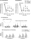
Figure 1
Figure 1
Attenuation of mechanical allodynia and neuronal hyperexcitability by administration of GABA. Spinal T13 hemisection injury results in bilateral mechanical allodynia on the contralateral (A, uninjured side) and the ipsilateral (B, injured side) hindlimbs compared to preinjury values before spinal hemisection (BH). On post operation day 28 (B i.t, before intrathecal administration), intrathecal 0.1 (#) and 0.5 (*) µg GABA administration significantly affects the mechanical allodynia in both hindlimbs compared to before intrathecal administration (p<0.05). Arrow reflects the time point of intrathecal administration. (C) displays the typical peri-stimulus histogram of lumbar wide dynamic range (WDR) dorsal horn neurons produced after GABA administration. After T13 hemisection, the lumbar WDR dorsal horn neurons show hyperexcitability (compare to uninjured, before SCI). Topical administration of GABA (arrow, 0.1 µg) significantly attenuated the hyperexcitability. (D) displays significant changes in evoked activity in the lumbar WDR dorsal horn neurons among the three different groups (p<0.05). The data reflect evoked activity at 30 min post injection (modified from Gwak et al., 2008).
Figure 2
Figure 2
Diagrams of inter- and intracellular mechanisms for the control of GABA following SCI. (A) In the normal condition, GABA concentrations are controlled by neuronal and glial GABA transporters and are dependent on the glutamate-GABA cycle. (B) After spinal cord injury, activated primary afferent fibers release huge concentration of GABA to prevent glutamate excitotoxicity. Elevation of extracellular GABA activates GABA transporters on neurons and activated glial cells. Once GABA is taken up, GABA is converted to glutamate and glutamine, which serves as a source of glutamate in neurons. This results in high concentrations of glutamate and GABA. In addition, glutamate and other pain stimulating agents, such as proinflammatory cytokines, ROS and ATP, released by activated glial cells, initiate activation of intracellular downstream events and modulate specific gene expression that result in downregulation of GAD expression. This positive feed forward cycle “GABA-glutamate-glutamine” contributes to hypofunction of GABA and persistent neuronal hyperexcitability, and plays a key role as a mechanism for neuropathic pain after SCI. Abbreviations; EAAs : excitatory amino acids, CREB and Elk : transcriptional factors, GAD : glutamic acid decarboxylase, GATs : GABA transporters, IL-1 : interleukin-1, IL-6 : interleukin-6, MAPK : mitogen activated protein kinase, NO : nitric oxide, PGs : prostaglandins, ROS : reactive oxygen species, SCI : spinal cord injury, TNF : tumor necrosis factor.
Figure 3
Figure 3
GABAergic excitation facilitates pain transmission in the spinal dorsal horn neurons following SCI. (A) After SCI, primary afferent fibers release excitatory neurotransmitters, such as glutamate, and initiate activation of glutamate (ionotropic and metabotropic) receptors. Massive influx of calcium ions trigger intracellular downstream pathways and contribute to neuronal excitation that results in enhanced pain transmission. (B) After SCI, upregulated NKCC1 induces influx of Cl into the cell that results in increased intracellular Cl. Activation (opened state) of the GABAA receptor facilitates efflux of Cl. Due to the Cl- gradient, the final net of Cl is efflux, producing membrane depolarizarion and enhancing the release of excitatory neurotransmitters, such as glutamate. (C) BDNF, released by activated microglia after SCI, activates trkB and contributes to the downregulation of KCC2. Inhibition of Cl efflux produces increased intracellular Cl concentrations. However, activation (opened state) of GABAA receptor facilitates a massive efflux of Cl and produces membrane depolarizarion that results in activation of voltage-dependent cation channels, such as Na+ and Ca2+ channels. This leads to neuronal hyperexcitability and neuropathic pain. Abbreviations; Glu : glutamate, iGluR : ionotropic glutamate receptor, mGluR : metabotropic glutamate receptor, BDNF : brain-derived neurotropic factor, NKCC1 : Na+-K+-Cl cotransporter 1, KCC2 : K+-Cl cotransporter 2, APs : action potentials, TrkB : tyrosine kinase receptor.

References

    1. Albrecht J, Pearce B, Murphy S. Evidence for an interaction between GABAB and glutamate receptors in astrocytes as revealed by changes in Ca2+ flux. Eur. J. Pharmacol. 1986;125:463–464. - PubMed
    1. Anderson CM, Swanson RA. Astrocyte glutamate transport: review of properties, regulation, and physiological functions. Glia. 2000;32:1–14. - PubMed
    1. Aptel H, Hilaire C, Pieraut S, Boukhaddaoui H, Mallie S, Valmier J, Scamps F. The Cav3.2/alpha1H T-type Ca2+ current is a molecular determinant of excitatory effects of GABA in adult sensory neurons. Mol. Cell. Neurosci. 2007;36:293–303. - PubMed
    1. Bao F, Liu D. Peroxynitrite generated in the rat spinal cord induces apoptotic cell death and activates caspase-3. Neuroscience. 2003;116:59–70. - PubMed
    1. Barakat L, Bordey A. GAT-1 and reversible GABA transport in Bergmann glia in slices. J. Neurophysiol. 2002;88:1407–1419. - PubMed

Publication types

MeSH terms

Substances