. 2011 Jan;23(1):333-49.
doi: 10.1105/tpc.110.078170. Epub 2011 Jan 7.

The nucleus-encoded trans-acting factor MCA1 plays a critical role in the regulation of cytochrome f synthesis in Chlamydomonas chloroplasts

Affiliations

The nucleus-encoded trans-acting factor MCA1 plays a critical role in the regulation of cytochrome f synthesis in Chlamydomonas chloroplasts

Alix Boulouis et al. Plant Cell. 2011 Jan.

Abstract

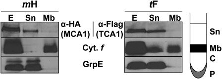
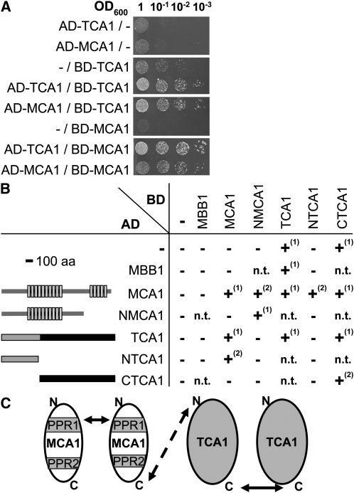
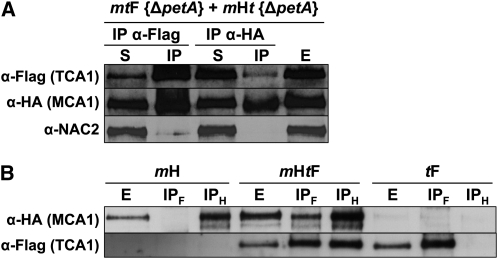
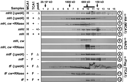
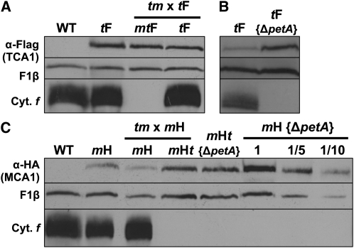
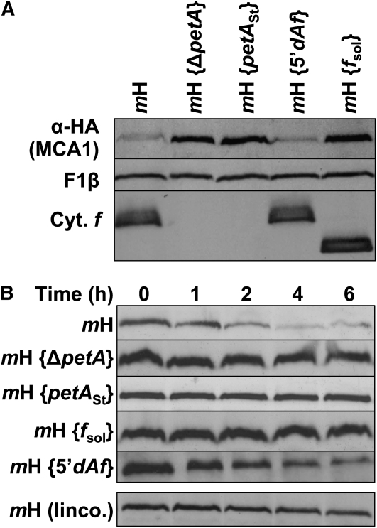
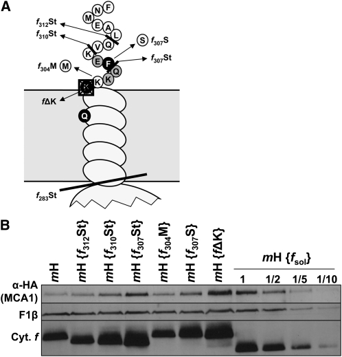
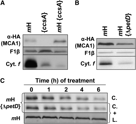
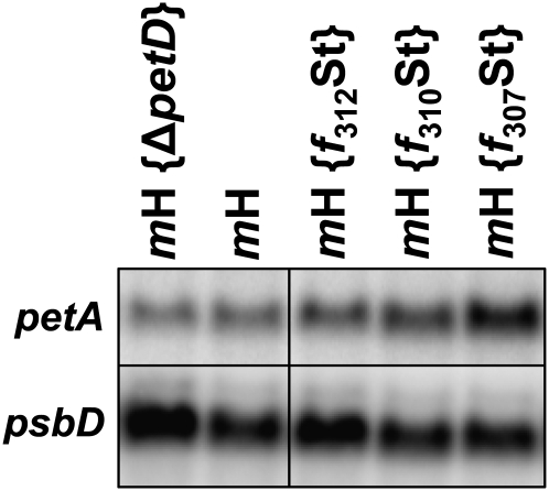
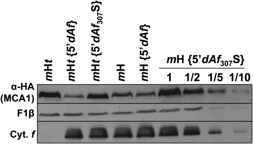
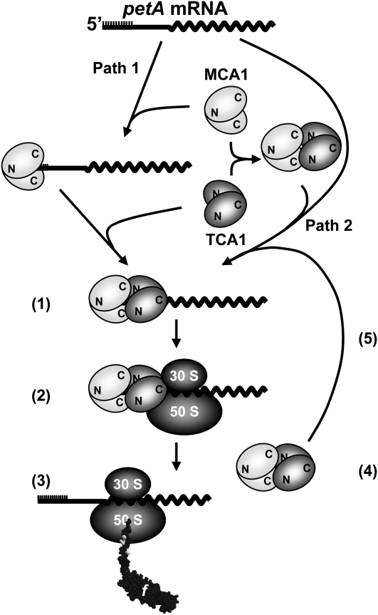
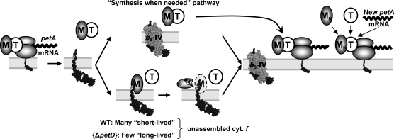
Organelle gene expression is characterized by nucleus-encoded trans-acting factors that control posttranscriptional steps in a gene-specific manner. As a typical example, in Chlamydomonas reinhardtii, expression of the chloroplast petA gene encoding cytochrome f, a major subunit of the cytochrome b(6)f complex, depends on MCA1 and TCA1, required for the accumulation and translation of the petA mRNA. Here, we show that these two proteins associate in high molecular mass complexes that also contain the petA mRNA. We demonstrate that MCA1 is degraded upon interaction with unassembled cytochrome f that transiently accumulates during the biogenesis of the cytochrome b(6)f complex. Strikingly, this interaction relies on the very same residues that form the repressor motif involved in the Control by Epistasy of cytochrome f Synthesis (CES), a negative feedback mechanism that downregulates cytochrome f synthesis when its assembly within the cytochrome b(6)f complex is compromised. Based on these new findings, we present a revised picture for the CES regulation of petA mRNA translation that involves proteolysis of the translation enhancer MCA1, triggered by its interaction with unassembled cytochrome f.


Figures

Figure 1.
Figure 1.
MCA1 and TCA1 Are Soluble Proteins. Cell extracts (E) from tF and mH strains, overlaid on a 1.5 M sucrose cushion, were separated by ultracentrifugation into the fractions schematically depicted in the right panel. Sn, supernatant; Mb, membrane; C, cushion; P, pellet. Equal volumes of each fraction were immunoreacted with anti-HA or anti-Flag antibodies. In addition, GrpE and cytochrome f were used as markers of soluble and membrane fractions, respectively. In the tF (TCA1) panel, fraction Sn was slightly contaminated by membranes, which explains the presence of cytochrome f (Cyt. f). Fractions C and P are not shown, as they were devoid of significant amounts of MCA1, TCA1, GrpE, or cytochrome f.
Figure 2.
Figure 2.
Interactions between MCA1 and TCA1 Probed by Two-Hybrid Experiments in Yeast. (A) Interactions between full-length MCA1 and TCA1 proteins. Interactions were assessed between the proteins indicated at the left of the figure, fused either to the AD or to the BD of the Gal4 transcription factor, by spotting serial dilutions of cotransformed yeast cells on selective medium (SD lacking Leu, Trp, and His but supplemented with 5 mM 3-AT). (B) Interactions observed between the truncated versions of MCA1 and TCA1 schematically depicted at the left of the figure. Light-gray boxes depict the PPR repeats in the MCA1 protein, while the thick black bar indicates the shortest region of TCA1 still able to complement tca1 mutations. Growth of transformed yeast was tested as above on different selective media: –, +(1), and +(2) indicate the absence of growth on any selective media, growth on medium lacking His and supplemented with 3-AT, and growth on a more stringent medium lacking His and adenine. n.t., not tested; aa, amino acids. (C) Schematic representation of MCA1/TCA1 interactions. PPR1 and 2 represent the two blocks of PPR repeats within MCA1. The dashed arrow points to an interaction suggested, but not fully demonstrated by our experiments, between the corresponding domains.
Figure 3.
Figure 3.
Interactions among MCA1, TCA1, and petA mRNA Probed by CoIP. (A) MCA1 and TCA1 interact in the absence of petA mRNA. Equal volumes of mtF {ΔpetA} and mHtpetA} cultures were mixed and broken with a French press. The soluble extract recovered after centrifugation (E) was immunoprecipitated with anti-HA or anti-Flag antibodies, as indicated. The presence of MCA1/TCA1 in supernatant (S) and immunoprecipitated (IP) fractions was then assessed using the same antibodies, as indicated at the left of the figure. The antibody against the NAC2 protein provided a specificity control. (B) Interactions between MCA1 and TCA1 in the presence of the petA mRNA. Soluble extracts (E) from strains mH, mHtF, and tF and immunoprecipitates recovered after incubation of the extracts with either anti-HA (IPH) or anti-Flag (IPF) antibodies were analyzed as above for the presence of MCA1 and TCA1.
Figure 4.
Figure 4.
Interactions between MCA1, TCA1, and the petA mRNA Probed by Size Exclusion Chromatography. Soluble or stromal (prepared from cw strains lacking cell wall) extracts from strains listed at the left of the figure were fractionated on Sephacryl S500 column and analyzed with the antibodies indicated at the right of the figure. Molecular masses of the complexes found in each fraction were estimated by comparison with standards of the HMW gel filtration calibration kit (GE Healthcare) or with the position of ribulose-1,5-bisphosphate carboxylase/oxygenase (indicated by the thick black bar at the top of the figure). The middle table recapitulates the status of each partner of the petA gene expression system: MCA1 (/TCA1) is absent (−) or expressed from the wild-type (+) or tagged (H/F) gene, while the petA mRNA is either absent in deletion strains (Δ) or in the absence of MCA1 (−), present in reduced amounts in the absence of TCA1 (+/−), or accumulated to wild-type levels (+).
Figure 5.
Figure 5.
The Accumulation of MCA1 or TCA1 Depends on the Presence of Their Partners from the petA Gene Expression System. (A) and (B) Immunoblots showing accumulation of TCA1-Fl in an otherwise wild-type genetic context (strain tF), in progeny expressing TCA1-Fl of one representative tetratype tetrad recovered after crossing mt and tF strains (A), or in the absence of petA mRNA (strain tF {ΔpetA}; [B]). The mtF progeny lacking MCA1 also lacks cytochrome f. (C) Accumulation of MCA1-HA in strains with an otherwise wild-type genetic context (mH), lacking petA (mH {ΔpetA}), both petA and TCA1 (mHtpetA}), and in the progeny expressing MCA1-HA of one representative tetratype tetrad from the cross mt × mH. The mHt progeny that inherited the mutant tca1 allele from the mt parent lacks cytochrome f. A dilution series of the mH {ΔpetA} sample is shown for the ease of quantification. For both panels, expression of cytochrome f (cyt. f) and of the β-subunit of the mitochondrial ATP synthase complex (F1β; loading control) is also shown. The wild-type strain (WT) that does not express MCA1-HA or TCA1-Fl is shown as a control.
Figure 6.
Figure 6.
Half-Life and Accumulation of MCA1 Are Governed by the Expression of Full-Length Cytochrome f. (A) Accumulation of MCA1-HA in an otherwise wild-type strain (mH), in strains carrying a petA gene deletion (mH {ΔpetA}) or expressing a modified petA gene that (1) cannot be translated (mH {petASt}), (2) is expressed under the control of the atpA 5′UTR (mH {5′dAf}), or (3) encodes a truncated soluble cytochrome f (mH {fSol}). Accumulation of cytochrome f (Cyt. f) in the various strains is shown, while the β-subunit of the mitochondrial ATP synthase complex (F1β) provides a loading control. (B) Half-life of MCA1 assessed in the same strains by immunochase at the various time points indicated after addition at t = 0 of cycloheximide alone or of cycloheximide plus lincomycin (linco.).
Figure 7.
Figure 7.
Disruption of the CES Repressor Domain Stabilizes MCA1. (A) Schematic representation of the mutations introduced in the stromal-exposed C-terminal tail of cytochrome f in strain mH. Substitutions of the residues shaded in black abolish the cytochrome f CES process, whereas mutations of those shaded in gray attenuate this regulatory process (Choquet et al., 2003). (B) Accumulation of MCA1-HA in strains with an otherwise wild-type background (strain mH) or carrying the mutations depicted in (A). A dilution series of the strain mH {fsol} is shown for the ease of quantification. Note that strain mH {fΔK} was slightly overloaded in this gel. Cyt., cytochrome.
Figure 8.
Figure 8.
Accumulation and Half-Life of MCA1 Are Governed by the Accumulation of the CES Repressor Motif. (A) Accumulation of MCA1-HA and cytochrome f (Cyt. f) in a double mutant mH, {ccsA-B6}, and in the parental strains mH and {ccsA-B6}. (B) Accumulation of MCA1-HA and cytochrome f in strains mH and mH {ΔpetD}. In both panels, the accumulation of the β-subunit of the mitochondrial ATP synthase complex (F1β) provides a loading control. (C) Half-life of MCA1-HA assessed in strains mH and mH {ΔpetD} by immunochase experiments in the presence of cycloheximide (C.) or both cycloheximide and lincomycin (C.+L.).
Figure 9.
Figure 9.
petA Transcript Accumulation in Representative Strains. Accumulation of petA mRNA in representative strains analyzed in Figures 7B and 8B. A probe specific for the psbD transcript provides a loading control. Note the overloading of sample mH {ΔpetD} and to a lesser extent of sample mH {f312St}.
Figure 10.
Figure 10.
Destabilization of MCA1 by the Cytochrome f C Terminus Does Not Require the Presence of TCA1. Accumulation of MCA1-HA, in an otherwise wild-type background (strain mH), in strains where petA coding sequence is expressed under the control of the atpA 5′UTR (strain mH {5′dAf}) and additionally carries a mutation preventing the CES process (strain mH {5′dAf307S}). A dilution series is shown for strain mH {5′dAf307S}. The same chloroplast genotypes were also compared in the mHt nuclear background (i.e., in the absence of TCA1). Accumulations of cytochrome f (Cyt. f) and subunit β of the mitochondrial ATP synthase (F1β, loading control) are also shown in the various strains.
Figure 11.
Figure 11.
Working Model of the Biogenesis of the petA Gene Expression System. MCA1 and TCA1 likely act in vivo as homo- and hetero-oligomers, whose formation would involve N- and C-terminal protein domains as indicated. MCA1 can bind to petA mRNA in the absence of TCA1 (Path 1) and also can interact with TCA1 in the absence of petA mRNA (Path 2). Both Paths 1 and 2 likely contribute in vivo to the formation of the ternary complex of ~600 kD (1) required for ribosome recruitment (2) and cytochrome f synthesis (3). Once translation is initiated, MCA1 and TCA1 would probably dissociate (4) from the translated mRNA and can be recycled for the formation of a new ternary complex (5) and new rounds of cytochrome f synthesis. Another interaction between MCA1 and unassembled cytochrome f is not shown in this figure but is detailed in Figure 12.
Figure 12.
Figure 12.
A Refined Model for the CES Process for Cytochrome f. The biogenesis of the cytochrome b6f complex in the wild type (WT) “synthesis when needed” pathway; see text. In strains with disrupted CES repressor domain, such as f307St, MCA1 (M) cannot interact with unassembled cytochrome (cyt.) f (f) and is efficiently recycled for new rounds of petA mRNA translation (top part of the figure). In the wild type, free C-terminal tails of cytochrome f accumulate transiently before assembly (“many short-lived” unassembled cytochrome f; bottom part of the figure) and may interact with some MCA1 proteins that are thus targeted for degradation. By contrast, in strains defective for cytochrome b6f complex assembly, such as ΔpetD, C-terminal tails of cytochrome f, although synthesized in reduced amount, are particularly long lived and would target most neighboring MCA1 for degradation (“few long-lived” unassembled cytochrome f; bottom part of the figure). The right side of the figure illustrates the formation of new ternary complexes following import of newly synthesized MCA1 (MN). De novo biogenesis of cytochrome f in assembly-defective cytochrome b6f mutants, such as {ΔpetD}, would mostly rely on these new ternary complexes. For the sake of clarity, MCA1 and TCA1 (T) are drawn as monomers, even if they are likely oligomeric in vivo (Figure 11).

References

    1. Ackerman S.H., Tzagoloff A. (2005). Function, structure, and biogenesis of mitochondrial ATP synthase. Prog. Nucleic Acid Res. Mol. Biol. 80: 95–133 - PubMed
    1. Anderson S., et al. (1981). Sequence and organization of the human mitochondrial genome. Nature 290: 457–465 - PubMed
    1. Auchincloss A.H., Zerges W., Perron K., Girard-Bascou J., Rochaix J.-D. (2002). Characterization of Tbc2, a nucleus-encoded factor specifically required for translation of the chloroplast psbC mRNA in Chlamydomonas reinhardtii. J. Cell Biol. 157: 953–962 - PMC - PubMed
    1. Balczun C., Bunse A., Hahn D., Bennoun P., Nickelsen J., Kück U. (2005). Two adjacent nuclear genes are required for functional complementation of a chloroplast trans-splicing mutant from Chlamydomonas reinhardtii. Plant J. 43: 636–648 - PubMed
    1. Barkan A., Goldschmidt-Clermont M. (2000). Participation of nuclear genes in chloroplast gene expression. Biochimie 82: 559–572 - PubMed

Publication types

MeSH terms