. 2011 Mar 25;286(12):10681-9.
doi: 10.1074/jbc.M110.196923. Epub 2011 Jan 7.

New insights into the signaling mechanism of the pH-responsive, membrane-integrated transcriptional activator CadC of Escherichia coli

Affiliations

New insights into the signaling mechanism of the pH-responsive, membrane-integrated transcriptional activator CadC of Escherichia coli

Ina Haneburger et al. J Biol Chem. .

Abstract

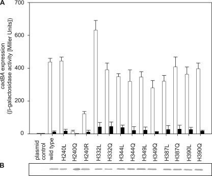
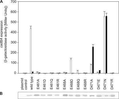
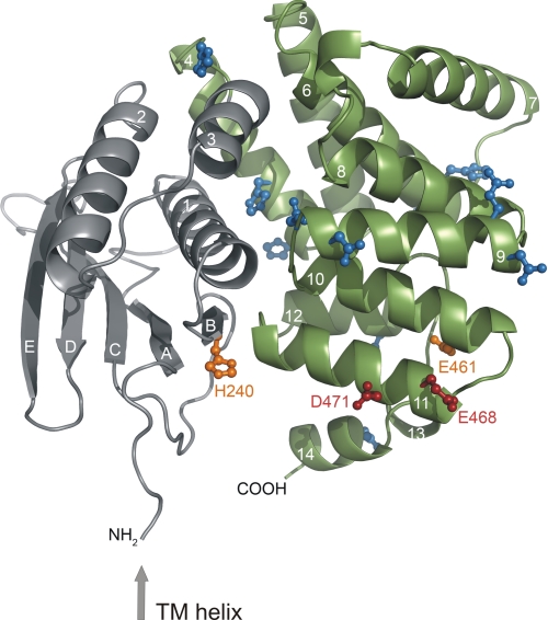
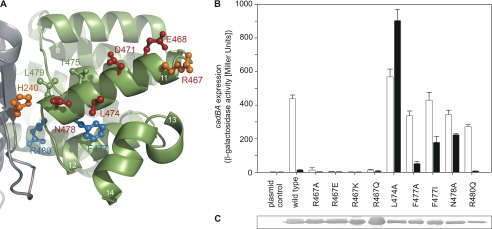
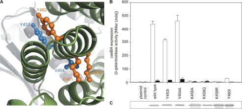
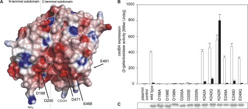
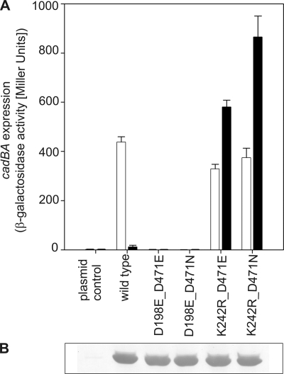
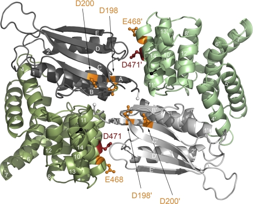
The membrane-integrated transcriptional regulator CadC of Escherichia coli activates expression of the cadBA operon at low external pH with concomitantly available lysine, providing adaptation to mild acidic stress. CadC is a representative of the ToxR-like proteins that combine sensory, signal transduction, and DNA-binding activities within a single polypeptide. Although several ToxR-like regulators such as CadC, as well as the main regulator of Vibrio cholerae virulence, ToxR itself, which activate gene expression at acidic pH, have been intensively investigated, their molecular activation mechanism is still unclear. In this study, a structure-guided mutational analysis was performed to elucidate the mechanism by which CadC detects acidification of the external milieu. Thus, a cluster of negatively charged amino acids (Asp-198, Asp-200, Glu-461, Glu-468, and Asp-471) was found to be crucial for pH detection. These amino acids form a negatively charged patch on the surface of the periplasmic domain of CadC that stretches across its two subdomains. The results of different combinations of amino acid replacements within this patch indicated that the N-terminal subdomain integrates and transduces the signals coming from both subdomains to the transmembrane domain. Alterations in the phospholipid composition did not influence pH-dependent cadBA expression, and therefore, interplay of the acidic surface patch with the negatively charged headgroups is unlikely. Models are discussed according to which protonation of these acidic amino acid side chains reduces repulsive forces between the two subdomains and/or between two monomers within a CadC dimer and thereby enables receptor activation upon lowering of the environmental pH.


Figures

FIGURE 1.
FIGURE 1.
A, influence of histidine replacements in the periplasmic domain of CadC on cadBA expression. Reporter gene assays were performed with E. coli EP314 (cadC1::Tn10, cadA′::lacZ) that was complemented with plasmid-encoded cadC or the mutant indicated. Cells were cultivated under microaerobic conditions in minimal medium in the presence of 10 mm lysine at pH 7.6 (black bars) or at pH 5.8 (white bars). The activity of the reporter enzyme β-galactosidase was determined according to Miller (40) and served as a measure for cadBA expression. The experiment was performed in triplicate, and error bars indicate S.D. B, verification of production and integration of CadC variants into the cytoplasmic membrane of E. coli. E. coli BL21(DE3)pLysS was transformed with plasmids encoding either wild-type or mutant cadC. Each lane contained 25 μg of membrane protein. CadC was detected by a monoclonal mouse antibody against the His tag and an alkaline phosphatase-coupled secondary antibody.
FIGURE 2.
FIGURE 2.
A, influence of replacements of acidic amino acids in the C-terminal region of the CadC periplasmic domain on cadBA expression. Reporter gene assays were performed with E. coli EP314 (cadC1::Tn10, cadA′::lacZ fusion) that was complemented with plasmid-encoded cadC or the indicated cadC mutant. Cells were cultivated under microaerobic conditions in minimal medium with 10 mm lysine at pH 7.6 (black bars) or at pH 5.8 (white bars). The reporter enzyme β-galactosidase was determined as described in the legend to Fig. 1. B, verification of production and integration of CadC variants into the cytoplasmic membrane of E. coli (see Fig. 1).
FIGURE 3.
FIGURE 3.
Localization of histidine, glutamate, and aspartate residues investigated in the first mutagenesis screen within the three-dimensional structure of the periplasmic domain of CadC (41). The preceding transmembrane (TM) helix is indicated by an arrow. The N-terminal subdomain of the periplasmic domain is colored gray, and the C-terminal subdomain is colored green. The side chains of residues that were mutated but did not alter the pH response of the corresponding CadC variants are colored blue. Amino acids whose mutagenesis suggested a role in pH sensing are indicated in orange (off-state) or red (on-state). Secondary structure elements are labeled with white letters (41).
FIGURE 4.
FIGURE 4.
Helix 11 of the periplasmic domain of CadC. A, amino acids of helix 11 that were investigated are colored blue (wild type-like phenotype), orange (off-state), or red (on-state). Side chains that are depicted in green were previously identified by random mutagenesis (Thr-475 and Leu-479) (10). The color code of the subdomains and the labeling of secondary structure elements is the same as described in the legend to Fig. 3. B, influence of amino acid replacements in helix 11 in the periplasmic domain of CadC on cadBA expression. Reporter gene assays were performed with E. coli EP314 (cadC1::Tn10, cadA′::lacZ fusion) that was complemented with plasmid-encoded cadC or the indicated cadC mutant as described in the legend to Fig. 1. C, verification of production and integration of CadC variants into the cytoplasmic membrane of E. coli BL21(DE3)pLysS.
FIGURE 5.
FIGURE 5.
Helix 10 of the periplasmic domain of CadC. A, amino acids of helix 10 of the periplasmic domain of CadC embrace helix 11. Amino acids that were investigated are colored blue (wild type-like phenotype) or orange (off-state). The color code of the subdomains and the labeling of secondary structure elements is the same as described in the legend to Fig. 3. B, influence of amino acid replacements in helix 10 in the periplasmic domain of CadC on cadBA expression. Reporter gene assays were performed with E. coli EP314 (cadC1::Tn10, cadA′::lacZ fusion) that was complemented with plasmid-encoded cadC or the indicated cadC mutant as described in the legend to Fig. 1. Black bars, pH 7.6; white bars, pH 5.8. C, verification of production and integration of CadC variants into the cytoplasmic membrane of E. coli (see Fig. 1).
FIGURE 6.
FIGURE 6.
Acidic residues from both subdomains form a contiguous negatively charged surface patch. A, electrostatic surface coloring of the periplasmic domain of CadC. Negatively charged surfaces are shown in red, whereas positively charged areas are shown in blue. The positions of Asp-198, Asp-200, Glu-461, Glu-468, and Asp-471 at the protein surface are indicated by arrows. The boundary between the two subdomains is indicated by a white dashed line. The N (NH2) and C (COOH) termini are indicated. B, influence of replacements of amino acids in the N-terminal subdomain on cadBA expression. Reporter gene assays were performed as described in the legend to Fig. 1. Black bars, pH 7.6; white bars, pH 5.8. C, verification of production and integration of CadC variants into the cytoplasmic membrane of E. coli.
FIGURE 7.
FIGURE 7.
Intramolecular complementation of signaling off- and on-state mutants. A, influence of different combinations of amino acid replacements in the two subdomains on cadBA expression. Reporter gene assays were performed with E. coli EP314 (cadC1::Tn10, cadA′::lacZ fusion) as described in the legend to Fig. 1. Black bars, pH 7.6; white bars, pH 5.8. B, verification of production and integration of CadC variants into the cytoplasmic membrane of E. coli.
FIGURE 8.
FIGURE 8.
Residues involved in pH sensing are located at the CadC periplasmic domain dimer interface. To distinguish the monomers, they are colored in dark and pale colors, respectively. Residues that are crucial for pH sensing are indicated in orange (off-state) or red (on-state). The N-terminal subdomains of the periplasmic domain are colored gray, and the C-terminal subdomains are colored green. The N termini, which are preceded by the transmembrane helices, are indicated by white spheres, whereas the C termini are indicated by black spheres.

References

    1. Bearson S., Bearson B., Foster J. W. (1997) FEMS Microbiol. Lett. 147, 173–180 - PubMed
    1. Auger E. A., Redding K. E., Plumb T., Childs L. C., Meng S. Y., Bennett G. N. (1989) Mol. Microbiol. 3, 609–620 - PubMed
    1. Soksawatmaekhin W., Kuraishi A., Sakata K., Kashiwagi K., Igarashi K. (2004) Mol. Microbiol. 51, 1401–1412 - PubMed
    1. Fritz G., Koller C., Burdack K., Tetsch L., Haneburger I., Jung K., Gerland U. (2009) J. Mol. Biol. 393, 272–286 - PubMed
    1. Meng S. Y., Bennett G. N. (1992) J. Bacteriol. 174, 2670–2678 - PMC - PubMed

Publication types

MeSH terms

Associated data

LinkOut - more resources