. 2011 Apr 8;286(14):12803-11.
doi: 10.1074/jbc.M110.179630. Epub 2011 Jan 7.

HSP70 natively and specifically associates with an N-terminal dermcidin-derived peptide that contains an HLA-A*03 antigenic epitope

Affiliations

HSP70 natively and specifically associates with an N-terminal dermcidin-derived peptide that contains an HLA-A*03 antigenic epitope

Pawel Stocki et al. J Biol Chem. .

Abstract

Tumor cells very often have elevated expression of HSP70, the anti-apoptotic properties of which contribute to overall tumor survival. Independent of its anti-apoptotic properties, HSP70 was also suggested to be involved in the antigen presentation process by chaperoning cytosolic peptides, thus protecting them from rapid degradation and securing the peptide pool for further processing. In this study, we identified a 33-amino acid N-terminal dermcidin (DCD)-derived peptide from the repertoire of in vivo HSP70-associated peptides isolated from a leukemic cell line, K562. The DCD peptide has been previously shown to be involved in tumorigenesis, to increase tumor survival rate, to improve tumor stress resistance, and to aid growth. We show that HSP70 is a specific binding partner for the DCD prosurvival peptide and define an ATP-dependent DCD-binding site (GNPCH). We also identify an HLA-A*03 antigenic epitope within the DCD peptide, which follows and partially overlaps the HSP70-binding site (CHEASAAQK). This study describes the interaction between HSP70 and the DCD-derived prosurvival peptide, an interaction that may direct the peptide toward antigen presentation and independently contribute to the prosurvival mechanism mediated by DCD.


Figures

FIGURE 1.
FIGURE 1.
Fragmentation spectrum of the N-terminal DCD-derived peptide isolated with HSP70. HSP70 was purified from the K562 cell line and fragmented on a Finnigan LTQ-FT mass spectrometer. *, the most intense peaks that correlated with the DCD sequence (Table 1). m/z is the mass-to-charge ratio of ions. The DCD-derived peptide sequence was determined by de novo sequencing to be YDPEAASAPGSGNPCHEASAAQKENAGEDPGLA (Table 1). Other HSP70-associated peptides were described previously by Stocki et al. (5).
FIGURE 2.
FIGURE 2.
DCD protein sequence and positions of the HSP70-associated N-terminal DCD-derived peptide, DSEP, and PIF. Shown is the DCD protein sequence (gi:16751921) and the sequences of DSEP and PIF previously published (20, 21, 25). The DCD protein is shown divided into the signal peptide (secretory signal sequence), DSEP (the reported prosurvival peptide; underlined) (20, 21), the propeptide (the DCD fragment localized between two reported functional peptides), and DCD-1 (the reported antimicrobial peptide; underlined) (17). PIF, a glycosylated N-terminal DCD-derived peptide, was suggested to be involved in the development of cachexia (27). Shown in boldface is the amino acid mismatch compared with the DCD sequence.
FIGURE 3.
FIGURE 3.
DCD expression at the protein and mRNA levels in the K562 cell line. A, expression of DCD at the mRNA level. Total RNA was isolated from either the K562 or CCRF-CEM (CCRF) cell line and subjected to reverse transcription and PCR. GAPDH was used as a control. For the K562 cell line, both DCD and GAPDH primers amplified products of the desired sizes, 322 and 240 bp, respectively. The CCRF-CEM cell line was shown not to express DCD. B, expression of DCD at the protein level. DCD expression was checked in the K562 cell lysate under reducing (R) conditions in the presence and absence of IAA with antibody against DCD (clone A-20). The arrows indicate the most intense bands: *, without IAA, ∼66 kDa; **, with IAA, ∼9 kDa. As a negative control, the CCRF-CEM cell lysate was run, and GAPDH was used as a loading control.
FIGURE 4.
FIGURE 4.
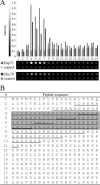
GNPCH is the DCD interaction site for HSP70. 19 peptides were derived from the DCD sequence (gi:16751921). The peptides were synthesized on a cellulose membrane. Peptides were 20 amino acids in length with a 15-amino acid fragment overlapping the previous peptide on the array. A, recombinant Hsp72 or Hsc70 was incubated with the array, followed by HRP-conjugated primary and secondary antibodies; a control was performed with the corresponding primary and secondary antibodies only. B, peptide sequences on the array. # indicates the peptide number on the array. The DCD peptide isolated with HSP70 is underlined. The spots on the array that were recognized by HSP70 are shaded. The 5-amino acid fragment (GNPCH) that was common for the peptides recognized by HSP70 is double-underlined.
FIGURE 5.
FIGURE 5.
Specific recognition of the DCD-derived peptide by HSP70 depends on the GNPCH fragment. Either the K562 whole cell lysate (A) or recombinant Hsp72 (B and C) was incubated with biotinylated peptides O, I, and S. C, Hsp72 was incubated with biotinylated N-terminal DCD-derived peptide O on its own or in the presence of 10 mm ATP, ADP, or ATPγS. The samples were run on a native gel, followed by transfer to nitrocellulose. Biotinylated peptide localization was visualized with HRP-conjugated streptavidin; Hsp72 was visualized with anti-HSP70 antibody as a loading control. D, the sequences of the biotinylated peptides used in the experiments are shown: peptide O, the DCD-derived peptide of original sequence as isolated from HSP70 (double-underlined), HSP70 recognition site; peptide I, the DCD peptide with an inverted HSP70 recognition site (underlined), the inverted fragment HCPNG; and peptide S, a scrambled peptide with the same residual abundance to peptide O but in a random sequence.
FIGURE 6.
FIGURE 6.
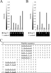
Alanine substitution analysis of the HSP70 interaction site within DCD. A, peptides designed to contain subsequent alanine substitutions in the fragment recognized by HSP70 (GNPCH) were synthesized on a peptide array and revealed the requirement of proline for efficient Hsp72 binding. B, two known antigenic peptides, Pmel-17/gp100 (peptide 7) and influenza A virus nucleoprotein-derived peptide (peptide 10), and their variants were immobilized on the membrane and tested for their affinity for Hsp72. The variants of these antigenic peptides had either an additional HSP70-binding motif on their N termini (GNPCH; peptides 8 and 11) or the modified non-recognizable motif (GNACH; peptides 9 and 12). Recombinant Hsp72 was incubated with the arrays, followed by HRP-conjugated primary and secondary antibodies; a control was performed with primary and secondary antibodies only. C, shown are peptide sequences that were synthesized on the peptide arrays. Double-underlined, HSP70 interaction site; underlined, alanine substitutions.
FIGURE 7.
FIGURE 7.
DCDp shows HLA-A*03 antigenic potential in vitro. A, purified HLA-A*03 T cells were subjected to stimulation with the KDI, DCDp, or Pmel-17/gp100 peptide. After six rounds of restimulation, CD8+ T cells were analyzed by FACS for the coexpression of the CD25 activation marker. The KDI, DCDp, and Pmel-17/gp100 peptides were tested for their ability to stimulate the proliferative T cell response (B) and IFNγ secretion (C). D, based on the SYFPEITHI algorithm, the KDI peptide was selected as a negative control; Pmel-17/gp100 was used as a stimulatory HLA-A*03 antigen. HLA-A*03 mo-iDCs were pulsed with 10 μm peptide overnight, washed, and used as stimulatory cells for the corresponding antigen-stimulated autologous T cells. The background T cell response to non-pulsed mo-iDCs was subtracted from the antigen-specific response.

References

    1. Peng P., Ménoret A., Srivastava P. K. (1997) J. Immunol. Methods 204, 13–21 - PubMed
    1. Srivastava P. (2002) Nat. Rev. Immunol. 2, 185–194 - PubMed
    1. Udono H., Srivastava P. K. (1993) J. Exp. Med. 178, 1391–1396 - PMC - PubMed
    1. Sato K., Torimoto Y., Tamura Y., Shindo M., Shinzaki H., Hirai K., Kohgo Y. (2001) Blood 98, 1852–1857 - PubMed
    1. Stocki P., Morris N. J., Preisinger C., Wang X. N., Kolch W., Multhoff G., Dickinson A. M. (2010) Cell Stress Chaperones 15, 729–741 - PMC - PubMed

Publication types