

Arginase 1 contributes to diminished coronary arteriolar dilation in patients with diabetes
- PMID: 21217072
- PMCID: PMC3064303
- DOI: 10.1152/ajpheart.00831.2010
Arginase 1 contributes to diminished coronary arteriolar dilation in patients with diabetes
Abstract
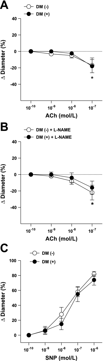
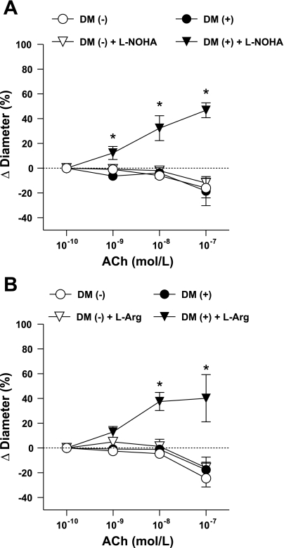
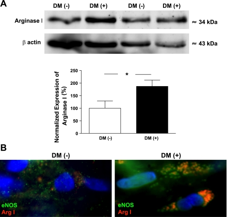
Arginase 1, via competing with nitric oxide (NO) synthase for the substrate L-arginine, may interfere with NO-mediated vascular responses. We tested the hypothesis that arginase 1 contributes to coronary vasomotor dysfunction in patients with diabetes mellitus (DM). Coronary arterioles were dissected from the right atrial appendages of 41 consecutive patients with or without DM (the 2 groups suffered from similar comorbidities), and agonist-induced changes in diameter were measured with videomicroscopy. We found that the endothelium-dependent agonist ACh elicited a diminished vasodilation and caused constriction to the highest ACh concentration (0.1 μM) with a similar magnitude in patients with (18 ± 8%) and without (17 ± 9%) DM. Responses to ACh were not significantly affected by the inhibition of NO synthesis with N(G)-nitro-L-arginine methyl ester in either group. The NO donor sodium nitroprusside-dependent dilations were not different in patients with or without DM. Interestingly, we found that the presence of N(G)-hydroxy-L-arginine (10 μM), a selective inhibitor of arginase or application of L-arginine (3 mM), restored ACh-induced coronary dilations only in patients with DM (to 47 ± 6% and to 40 ± 19%, respectively) but not in subjects without DM. Correspondingly, the protein expression of arginase 1 was increased in coronary arterioles of patients with DM compared with subjects without diabetes. Moreover, using immunocytochemistry, we detected an abundant immunostaining of arginase 1 in coronary endothelial cells of patients with DM, which was colocalized with NO synthase. Collectively, we provided evidence for a distinct upregulation of arginase 1 in coronary arterioles of patients with DM, which contributes to a reduced NO production and consequently diminished vasodilation.
Figures
References
-
- Bagi Z, Koller A. Lack of NO-mediation of flow-dependent arteriolar dilation in diabetes is restored by sepiapterin. J Vasc Res 40: 47–57, 2003 - PubMed
-
- Bagi Z, Koller A, K G. Peroxisome proliferator-activated receptor-γ activation, by reducing oxidative stress Increases NO bioavailability in coronary arterioles in type 2 diabetes. Am J Physiol Heart Circ Physiol 286: H742–H748, 2004 - PubMed
-
- Beckman JA, Goldfine AB, Gordon MB, Creager MA. Ascorbate restores endothelium-dependent vasodilation impaired by acute hyperglycemia in humans. Circulation 103: 1618–1623, 2001 - PubMed
-
- Cannon RO, 3rd, Camici PG, Epstein SE. Pathophysiological dilemma of syndrome X. Circulation 85: 883–892, 1992 - PubMed
Publication types
MeSH terms
Substances
Grants and funding
LinkOut - more resources
Full Text Sources
Other Literature Sources
Medical
Research Materials