. 2011 Mar 10;117(10):2901-9.
doi: 10.1182/blood-2010-05-286351. Epub 2011 Jan 7.

Notch is oncogenic dominant in T-cell acute lymphoblastic leukemia

Affiliations

Notch is oncogenic dominant in T-cell acute lymphoblastic leukemia

Renée M Demarest et al. Blood. .

Erratum in

  • Blood. 2011 Jun 16;117(24):6739

Abstract

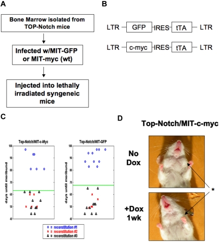
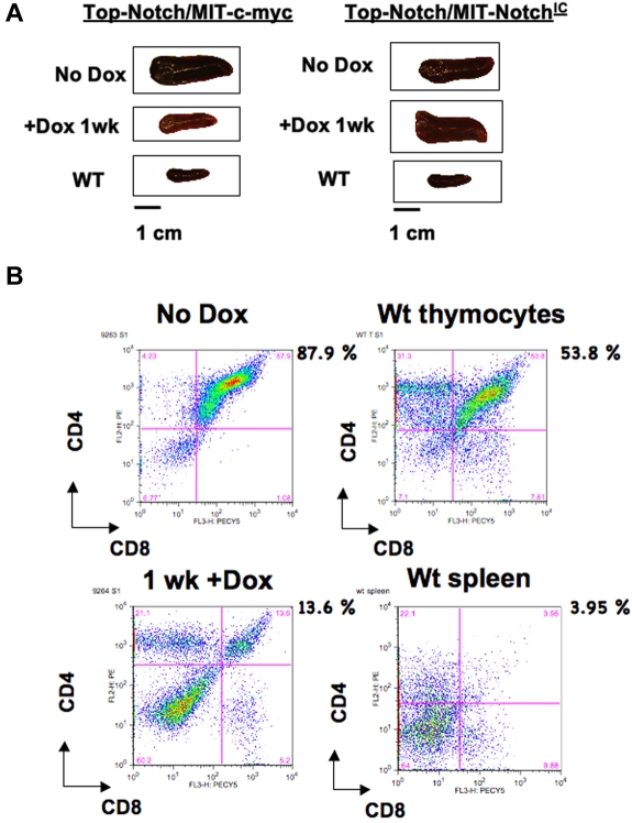
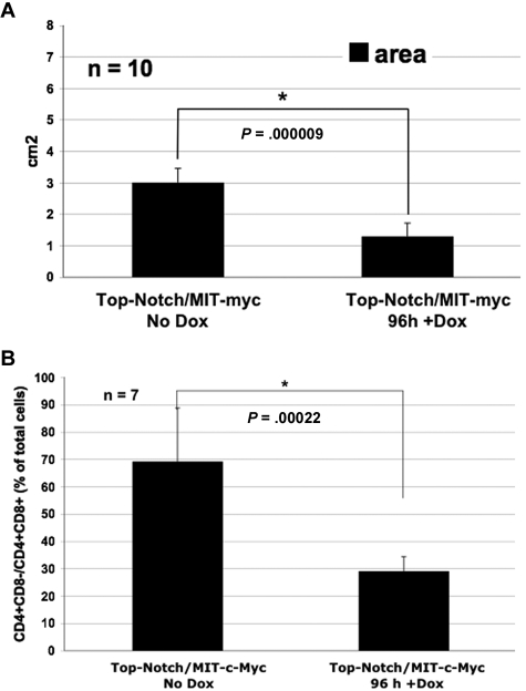
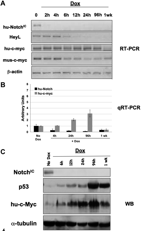
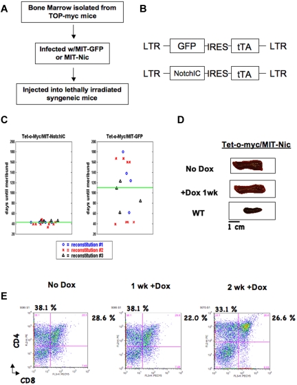
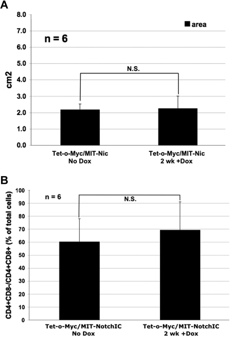
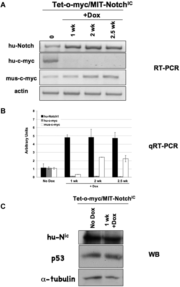
T-cell acute lymphoblastic leukemia (T-ALL) is a hematologic neoplasm characterized by malignant expansion of immature T cells. Activated NOTCH (Notch(IC)) and c-MYC expression are increased in a large percentage of human T-ALL tumors. Furthermore, c-MYC has been shown to be a NOTCH target gene. Although activating mutations of Notch have been found in human T-ALL tumors, there is little evidence that the c-MYC locus is altered in this neoplasm. It was previously demonstrated that Notch and c-Myc-regulated genes have a broadly overlapping profile, including genes involved in cell cycle progression and metabolism. Given that Notch and c-Myc appear to function similarly in T-ALL, we sought to determine whether these two oncogenes could substitute for each other in T-ALL tumors. Here we report that NOTCH(IC) is able to maintain T-ALL tumors formed in the presence of exogenous NOTCH(IC) and c-MYC when exogenous c-MYC expression is extinguished. In contrast, c-MYC is incapable of maintaining these tumors in the absence of NOTCH(IC). We propose that failure of c-MYC to maintain these tumors is the result of p53-mediated apoptosis. These results demonstrate that T-ALL maintenance is dependent on NOTCH(IC), but not c-MYC, demonstrating that NOTCH is oncogenic dominant in T-ALL tumors.


Figures

Figure 1
Figure 1
Coexpression of ectopic c-MYC does not cooperate in Top-NOTCH tumor formation or prevent tumor regression. (A) Schematic of the tumor formation protocol. (B) Diagram of retroviral constructs. (C) Death curve of mice reconstituted with Top-NOTCH bone marrow infected with MIT-c-MYC (left panel) and MIT-GFP (right panel). (D) Top-NOTCH/MIT-c-MYC mouse before and after treatment with Dox. *Reduction of lymph nodes.
Figure 2
Figure 2
T-ALL tumors are not maintained by c-MYC in the absence of NOTCHIC. (A) Spleens from Top-NOTCH/MIT-c-MYC (left panel) and Top-NOTCH/MIT-NOTCHIC (right panel) tumors treated for the indicated times with Dox. (B) FACS analysis using anti-CD4-PE and anti-CD8-PerCP-Cy5.5 antibodies to detect double-positive tumor cells in the spleen before and after Dox treatment of mice reconstituted with Top-NOTCH/MIT-c-MYC bone marrow (upper and lower left panels, respectively), wild-type (WT) thymus, and wild-type spleen (upper and lower right panels, respectively).
Figure 3
Figure 3
Spleen size of moribund mice reconstituted with Top-NOTCH/MIT-c-MYC bone marrow is significantly reduced after Dox treatment. (A) Area (length × width) of moribund mice reconstituted with Top-NOTCH/MIT-c-MYC bone marrow, untreated with Dox (No Dox) or treated for 96 hours with Dox. *P = .000009 (Student t test). n = 10. (B) Quantification of CD4+CD8 and CD4+CD8+ T cells in spleens of mice with Top-NOTCH/MIT-c-MYC tumors untreated or treated with Dox for 96 hours. *P = .00022 (Student t test). n = 7.
Figure 4
Figure 4
Expression of p53 is induced in T-ALL tumors when NOTCHIC expression is extinguished. Each primary tumor was transplanted into multiple syngeneic mice, and tumors were allowed to form. When mice were moribund, Dox was added and tissue collected at various time points for analysis. RT-PCR (A), quantitative RT-PCR (B), and Western blot (C) analyses were performed with the indicated specific primers or antibodies on single-cell suspensions from spleens of Top-NOTCHIC-/MIT-c-MYC tumors.
Figure 5
Figure 5
NOTCHIC expression can maintain Tet-o-MYC/MIT-NOTCHIC T-ALL tumors. (A) Schematic for generation of Tet-o-MYC/MIT-NOTCHIC tumors. (B) Diagram of retroviral constructs. (C) Death curves for mice reconstituted with Tet-o-MYC bone marrow infected with MIT-NOTCHIC (left panel) and MIT-GFP (right panel). (D) Spleens from nonregressing Tet-o-MYC/MIT-NOTCHIC. (E) FACS analysis using α-CD4-PE and α-CD8-PerCP-Cy5.5 antibodies on single-cell suspensions from spleens of moribund nonregressing Tet-o-MYC/MIT-NOTCHIC treated with Dox for the indicated times.
Figure 6
Figure 6
Spleen size of moribund mice reconstituted with Tet-o-MYC/MIT-NOTCHIC bone marrow does not significantly reduce after Dox treatment. (A) Area (length × width) of moribund mice reconstituted with Tet-o-MYC/MIT-NOTCHIC bone marrow, untreated with Dox (No Dox) or treated for 2 weeks with Dox. N.S. indicates not significant. P = .8257 (Student t test). n = 6. (B) Quantification of CD4+CD8 and CD4+CD8+ T cells in spleens of mice with Tet-o-MYC/MIT-NOTCHIC tumors untreated or treated with Dox for 2 weeks. There was no significant change (N.S.) in the size of spleens of mice with Tet-o-MYC/MIT-NOTCHIC treated with Dox (P = .8574; Student t test). n = 6.
Figure 7
Figure 7
NOTCHIC expression is maintained in nonregressing Tet-o-MYC/MIT-NOTCHIC tumors treated with Dox. (A) RT-PCR analysis of spleens from nonregressing Tet-o-MYC/MIT-NOTCHIC tumors treated with Dox for the indicated times using the indicated specific primers. (B) Quantitative RT-PCR analysis on single-cell suspensions from spleens of nonregressing Tet-o-MYC/MIT-NOTCHIC tumors treated with Dox. (C) Western blot analysis with antibodies against the indicated proteins in nonregressing Tet-o-MYC/MIT-NOTCHIC tumors treated with Dox.

Comment in

  • Notch, a T-ALL order.
    Osborne BA. Osborne BA. Blood. 2011 Mar 10;117(10):2749-50. doi: 10.1182/blood-2011-01-330225. Blood. 2011. PMID: 21393498 No abstract available.

References

    1. Goldberg JM, Silverman LB, Levy DE, et al. Childhood T-cell acute lymphoblastic leukemia: the Dana-Farber Cancer Institute acute lymphoblastic leukemia consortium experience. J Clin Oncol. 2003;21(19):3616–3622. - PubMed
    1. Pui CH, Sandlund JT, Pei D, et al. Improved outcome for children with acute lymphoblastic leukemia: results of Total Therapy Study XIIIB at St Jude Children's Research Hospital. Blood. 2004;104(9):2690–2696. - PubMed
    1. Thiel E, Kranz BR, Raghavachar A, et al. Prethymic phenotype and genotype of pre-T (CD7+/ER−)-cell leukemia and its clinical significance within adult acute lymphoblastic leukemia. Blood. 1989;73(5):1247–1258. - PubMed
    1. Grabher C, von Boehmer H, Look AT. Notch 1 activation in the molecular pathogenesis of T-cell acute lymphoblastic leukaemia. Nat Rev Cancer. 2006;6(5):347–359. - PubMed
    1. Aifantis I, Raetz E, Buonamici S. Molecular pathogenesis of T-cell leukaemia and lymphoma. Nat Rev Immunol. 2008;8(5):380–390. - PubMed

Publication types

Substances