. 2011 Feb 2;30(3):456-67.
doi: 10.1038/emboj.2010.348. Epub 2011 Jan 7.

Molecular mechanism of Ena/VASP-mediated actin-filament elongation

Affiliations

Molecular mechanism of Ena/VASP-mediated actin-filament elongation

Dennis Breitsprecher et al. EMBO J. .

Abstract

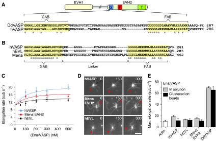
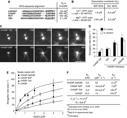
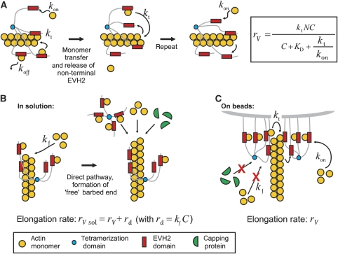
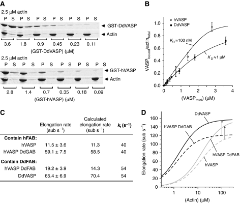
Ena/VASP proteins are implicated in a variety of fundamental cellular processes including axon guidance and cell migration. In vitro, they enhance elongation of actin filaments, but at rates differing in nearly an order of magnitude according to species, raising questions about the molecular determinants of rate control. Chimeras from fast and slow elongating VASP proteins were generated and their ability to promote actin polymerization and to bind G-actin was assessed. By in vitro TIRF microscopy as well as thermodynamic and kinetic analyses, we show that the velocity of VASP-mediated filament elongation depends on G-actin recruitment by the WASP homology 2 motif. Comparison of the experimentally observed elongation rates with a quantitative mathematical model moreover revealed that Ena/VASP-mediated filament elongation displays a saturation dependence on the actin monomer concentration, implying that Ena/VASP proteins, independent of species, are fully saturated with actin in vivo and generally act as potent filament elongators. Moreover, our data showed that spontaneous addition of monomers does not occur during processive VASP-mediated filament elongation on surfaces, suggesting that most filament formation in cells is actively controlled.


Conflict of interest statement

The authors declare that they have no conflict of interest.

Figures

Figure 1
Figure 1
Effects of hVASP, Mena and hEVL on actin-filament elongation. (A) Domain organization of Ena/VASP proteins and sequence alignment of the corresponding GAB-linker-FAB region within the EVH2 domains of DdVASP and hVASP. The GAB and FAB are highlighted in yellow. Identical amino acids within these motifs are marked with an asterisk. G, GAB; F, FAB; T, tetramerization domain. (B) Sequence alignment of the GAB-linker-FAB region of hVASP, hEVL and Mena. The linker length differs in all three proteins. (C) Elongation rates of 1.3 μM actin (30% Oregon-Green (OG) labelled) in the presence of different concentrations of hVASP, Mena EVH2 and hEVL determined by single-filament TIRFM in TIRF buffer (see Materials and methods). (D) TIRFM micrographs of the assembly of 1.3 μM actin (30% OG labelled) on beads saturated with hVASP, Mena EVH2 and hEVL in the presence of 80 nM CP. Arrows indicate growing filaments. Time is indicated in seconds, scale=10 μm. (E) Comparison of the maximal elongation rates of 1.3 μM actin (30% OG labelled) for the three mammalian Ena/VASP paralogues and DdVASP determined by TIRFM in solution or immobilized on beads. Elongation rates are presented as mean values±s.e.m.
Figure 2
Figure 2
Replacement of the GAB and FAB in hVASP with the corresponding DdVASP motifs markedly accelerates VASP-mediated filament elongation. (A) Scheme of hVASP chimeras bearing different domains of DdVASP. DdVASP is shown in colour and hVASP is shown in greyscale. G, GAB; F, FAB; T, tetramerization domain. (B) TIRFM micrographs of the assembly of 1.3 μM actin (30% OG labelled) in TIRF buffer containing 500 nM of the chimera are indicated. Time is indicated in seconds, scale=10 μm. (C) Elongation rates of the chimeras in solution in a concentration rage from 25 nM to 1 μM. (D) (Left) TIRFM micrographs of the assembly of 1.3 μM actin (30% OG labelled) in TIRF buffer in the presence of 200 nM CP on beads saturated with the hVASP chimeras are indicated. Time is shown in seconds, scale=10 μm. (Right) Plots of the lengths of individual filaments versus time yield filament elongation rates. Elongation rates are presented as mean values±s.e.m.
Figure 3
Figure 3
Actin-binding properties of the GAB motifs and EVH2 domains of hVASP and DdVASP. (A) Determination of the KD value of the DdGAB–actin interaction by fluorescence titration of 100 nM OG-actin (left) and 500 nM OG-actin (right) with the DdGAB peptide at the buffer conditions is indicated (for details see Materials and methods section). The solid lines represent calculated binding isotherms. (B) Determination of the KD value of the hGAB–actin interaction by fluorescence titration of 300 nM OG-actin (left) and 3 μM OG-actin (right) with the hGAB peptide at conditions as in (A). The solid lines represent calculated binding isotherms. (C) Summary of KD values determined from experiments similar as shown in (A, B) at the conditions indicated. KD values for the GAB–Ca2+–ATP–actin interaction are given in brackets. KD values obtained under conditions used in TIRF assays (50 mM KCl, Mg2+–ATP–actin) are shown in bold. (D) (Left) Kinetics of the DdEVH2–G-actin interaction. Time course of the binding of DdEVH2 to latA-sequestered, Mg2+–ATP–OG-actin was monitored by a stopped-flow apparatus at a final actin concentration of 2.5 μM actin and the DdEVH2 concentrations are indicated. Noisy curves represent experimental data, and solid lines are fits for a reversible, bimolecular reaction with KD=0.6 μM, yielding kon=53 μM−1 s−1. (Right) kon values of the interaction of the hEVH2 and DdEVH2 domain with OG-actin. Due to the low affinity of the hGAB, high actin concentrations had to be used (10 μM final), which resulted in more noisy signals (not shown) that could not be fitted as accurately as those for the higher-affinity DdEVH2.
Figure 4
Figure 4
Analysis of hVASP WH2 chimeras reveals VASP-mediated filament elongation is enhanced by saturable monomer binding to GAB sites. (A) Sequence alignment of the WH2 motifs is indicated. In the chimeric proteins, the GAB of hVASP was replaced by the WH2 motifs from Tβ4 and WIP. KD values of the actin–WH2 interaction in G-buffer where either determined in this study (*) or by Chereau et al (2005) (#). Conserved hydrophobic residues are highlighted in grey and the conserved LxxV/T motifs (x=basic amino acid) are boxed. (B) KD values determined as for Figure 3A and B under the conditions are indicated. (*) The KD values for the Tβ4–WH2 interaction were estimated on the basis of previous studies (De La Cruz et al, 2000; Hertzog et al, 2002; Chereau et al, 2005). (C) TIRFM micrographs of the assembly of 1.3 μM actin (30% OG labelled) on beads saturated with hVASP Tβ4 and hVASP WIP in TIRF buffer in the presence of 80 nM CP. Both chimeras processively elongate actin filaments. Time is indicated in seconds, scale=20 μm. (D) Elongation rates mediated by hVASP and the chimeras hVASP Tβ4, hVASP WIP and hVASP DdGAB at 1.3 μM G-actin either with 500 nM of the VASP constructs in solution or in the presence of 80 nM CP on saturated beads. Number of analysed filaments >20 for bead assays and >40 for assays in solution. Elongation rates are presented as mean values±s.e.m. (E) Elongation rates obtained from TIRF assays with beads coated with different hVASP constructs in the presence of 80 nM CP at the actin concentrations indicated. Solid lines represent best fits of the experimental data using the mathematical model for processive filament elongation by immobilized VASP as described in Figure 5 and the Materials and methods section. Elongation rates are presented as mean values±s.e.m. (F) Parameters derived from fitting of the data are shown in (E).
Figure 5
Figure 5
Mathematical model of VASP-mediated actin-filament elongation. (A) General mechanism of VASP-mediated filament elongation. A VASP tetramer is attached to the filament barbed end by at least one EVH2 domain during filament elongation. The free EVH2 domains recruit actin monomers from solution with an on-rate constant kon, and subsequently either transfer the subunit onto the barbed end with a transfer rate kt or release the actin monomer back into solution with an off-rate koff. This model also assumes that upon binding of the GAB-associated monomer to the barbed end, the already bound GAB is quickly (or simultaneously) released from the now penultimate subunit so that it becomes immediately available to capture another monomer. By this cycle, the VASP tetramer is able to processively track the elongating filament tip while cyclically maintaining one GAB bound to the terminal subunit and three GABs free to capture monomers from solution. (B) VASP-mediated actin-filament elongation in solution. When VASP is free in solution, both, processive association of VASP with the barbed end and spontaneous addition of free actin monomers via the direct pathway with an independent transfer rate constant kf can occur. Free barbed ends produced by the direct pathway are accessible either for other VASP tetramers, actin monomers or, if present, capping proteins. The average elongation rate is therefore the sum of the VASP-mediated filament elongation rate and the rate of the direct pathway. (C) VASP-mediated filament elongation on surfaces. Upon clustering, the high density of VASP at the surface leads to processive association of VASP with the filament end, efficiently blocking binding of capping proteins and also preventing spontaneous addition of monomers via the direct pathway. Hence, filament elongation is exclusively fueled by actin monomers recruited and transferred by VASP.
Figure 6
Figure 6
Function of the FAB motif in VASP-mediated filament elongation. (A) High-speed co-sedimentation analysis on the binding of GST-hVASP and GST-DdVASP to F-actin. In all, 2.5 μM actin was polymerized in polymerization buffer supplemented with VASP at the concentrations indicated. After centrifugation at 180 000 g for 30 min, supernatants and pellets were analysed by SDS–PAGE. (B) KD values of the VASP–F-actin interaction were estimated from densiometric analyses of the amounts of GST-VASP constructs in the pellets (bound VASP) and supernatants (free VASP) from experiments as shown in (A), and by assuming that VASP binds to F-actin in a 1:1 ratio (Samarin et al, 2003). Error bars represent s.e.m. from three experiments. (C) Potential role of the F-actin affinity of the FAB on VASP-mediated filament elongation. Taking into account that different FAB motifs might alter kt depending on their F-actin affinity, experimental data could be fitted with fixed KD and kon values, yielding a higher kt value for constructs bearing the DdFAB motif (kt values are highlighted in bold). Elongation rates were determined by TIRF microscopy and are presented as mean values±s.e.m. (D) Calculated effect of higher kt values on VASP-mediated filament elongation. Dashed lines represent calculated values for VASP constructs bearing the higher-affinity hFAB motif using kt=40 s−1. Solid lines represent calculated values for constructs with the lower-affinity DdFAB using kt=54 s−1. The arrows indicate that the faster kt increases the maximal elongation rate of VASP-mediated actin assembly by ∼30% under saturating conditions.
Figure 7
Figure 7
Calculation of the saturation dependence of filament elongation by Ena/VASP proteins. Comparison of the calculated elongation rates for processive filament elongation mediated by hVASP WT and hVASP DdGAB over a large range of actin concentrations. Grey boxes indicate G-actin concentrations typically used in TIRF assays or present in biomimetic motility assays (Samarin et al, 2003), as well as physiological G-actin concentrations, for instance in the lamellipodium tip (Koestler et al, 2009). Different G-actin affinities of the GAB motifs result in markedly different elongation rates mediated by chimeras hVASP and hVASP DdGAB in TIRF assays. However, both proteins are expected to be saturated with actin monomers and to enhance filament elongation to the same extent at very high monomer concentrations, for example under in vivo conditions.

References

    1. Ahuja R, Pinyol R, Reichenbach N, Custer L, Klingensmith J, Kessels MM, Qualmann B (2007) Cordon-bleu is an actin nucleation factor and controls neuronal morphology. Cell 131: 337–350 - PMC - PubMed
    1. Applewhite DA, Barzik M, Kojima S, Svitkina TM, Gertler FB, Borisy GG (2007) Ena/VASP proteins have an anti-capping independent function in filopodia formation. Mol Biol Cell 18: 2579–2591 - PMC - PubMed
    1. Bachmann C, Fischer L, Walter U, Reinhard M (1999) The EVH2 domain of the vasodilator-stimulated phosphoprotein mediates tetramerization, F-actin binding, and actin bundle formation. J Biol Chem 274: 23549–23557 - PubMed
    1. Barzik M, Kotova TI, Higgs HN, Hazelwood L, Hanein D, Gertler FB, Schafer DA (2005) Ena/VASP proteins enhance actin polymerization in the presence of barbed end capping proteins. J Biol Chem 280: 28653–28662 - PMC - PubMed
    1. Bear JE, Loureiro JJ, Libova I, Fässler R, Wehland J, Gertler FB (2000) Negative regulation of fibroblast motility by Ena/VASP proteins. Cell 101: 717–728 - PubMed

Publication types