. 2011 Feb;8(2):139-42.
doi: 10.1038/nmeth.1552. Epub 2011 Jan 9.

Simultaneous two-photon calcium imaging at different depths with spatiotemporal multiplexing

Affiliations

Simultaneous two-photon calcium imaging at different depths with spatiotemporal multiplexing

Adrian Cheng et al. Nat Methods. 2011 Feb.

Abstract

In vivo two-photon calcium imaging would benefit from the use of multiple excitation beams to increase scanning speed, signal-to-noise ratio and field of view or to image different axial planes simultaneously. Using spatiotemporal multiplexing we circumvented light-scattering ambiguity inherent to deep-tissue multifocal two-photon microscopy. We demonstrate calcium imaging at multiple axial planes in the intact mouse brain to monitor network activity of ensembles of cortical neurons in three spatial dimensions.


Conflict of interest statement

COMPETING FINANCIAL INTERESTS

The authors declare no competing financial interest

Figures

Figure 1
Figure 1
Spatiotemporal multiplexing to overcome depth limitations in multifocal 2PLSM. (a) Layout of the prototype microscope. Laser pulses are emitted with 12 ns period from a commercial ultrafast Ti:Al203 laser. The beam is divided into 4 beams, which are delayed by 3 ns each (1 m per 3 ns) and converged on the slow-axis scan mirror aperture, which is then projected onto the objective back aperture. The resulting emitted fluorescence, which is highly scattered, is collected by two hybrid photodetectors (HPDs). The HPD active area is placed in a demagnified conjugate plane of the objective back aperture to maximize scattered light collection. (b) Illustration of different beam scanning patterns at the sample. Time multiplexing removes ambiguity between different imaging planes, allowing both axial and lateral beam distribution. (c) Time course of detected fluorescence signal for a single beam (top) and four spatiotemporally multiplexed beams (bottom). Overlay of 200 oscilloscope traces and summary histograms of single photoelectron events (using a pollen grain). Fluorescence from different time windows (different colors) is associated with different delayed excitation beams. Scale bar: 12 ns.
Figure 2
Figure 2
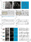
Multifocal 2-D and 3-D in vivo 2-photon calcium imaging (2PCI) of L2/3 neurons in barrel cortex with spatiotemporal multiplexing. (a) Spatial distribution of four beams in a single image plane (left) and typical field of view (right). The image is an average intensity time projection of a representative calcium imaging movie (3 min, 250 frames per second) from a P20 mouse using Fluo-4AM. Scale bar: 50 μm.(b) Zero-lag cross-correlation image computed from the movie (see Online Methods). (c) Final segmented image of cell bodies obtained through morphological filters (red contours). (d) Raw calcium traces of 11 different cells (neuropil signal in blue). Scale bar: 20 s.(e) Model calcium traces with identified neuronal spiking events (tic marks) of selected cells using a peeling algorithm (see Online Methods). Scalebar: 20 s. (f, g) Details of shaded regions shown in d and e, respectively. Scale bar: 5 s. (h) Spatial distribution of four beams arranged axially (left) and field of view for each imaging plane (right). Images are average intensity time projections of a typical movie with Fluo-4 AM (3 min, 60 volumes per second) with depth spanning from 90 μm to 180 μm below the pia (encompassing L1 to L3). Scale bar: 50 μm. (i) Zero-lag cross-correlation image (left) and fully segmented image (right) with cell contours (red). (j) Selected traces reconstructed by the peeling algorithm (right). Scale bar: 20 s.
Figure 3
Figure 3
Multifocal 2PCI with spatiotemporal multiplexing to assess activity-derived neuronal connectivity in L2/3 of barrel cortex. (a) Spatial distribution of four scanning beams (left) and representative field of view in two separate imaging planes (right) from an experiment with a P20 mouse using Fluo-4 AM (3 min, 100 frames per second). Scale bar: 50 μm. (b) Zero-lag cross-correlation image (left) and segmented image (right). (c) Raster plot showing identified spiking events in cells from b. Events shown in red were identified as having participated in a peak of synchrony (bottom trace). Scale bar: 20s.(d) Peak correlation coefficient (over a time lag of ± 1 s) for significantly correlated cells from b. (e) Axial (depth) versus radial (lateral) spread of bursts of neuronal firing corresponding to peaks of synchrony identified in several movies (n = 10 movies from 2 mice, n = 173 peaks of synchrony). A minority of bursts had a spatial organization consistent with either columnar (upper left) or laminar connectivity (bottom right). (f) Peak correlation coefficients from c versus cell pair radial distance (ΔR), for cell pairs in different imaging planes, the same imaging plane, and for all pairs (n = 10 movies, N = 10,262 pairs).

References

    1. Grewe BF, Helmchen F. Optical probing of neuronal ensemble activity. Curr Opin Neurobiol. 2009 doi: 10.1016/j.conb.2009.09.003. S0959-4388(09)00125-1 [pii] - DOI - PubMed
    1. Svoboda K, Yasuda R. Principles of two-photon excitation microscopy and its applications to neuroscience. Neuron. 2006;50:823–839. - PubMed
    1. Kerlin AM, Andermann ML, Berezovskii VK, Reid RC. Broadly tuned response properties of diverse inhibitory neuron subtypes in mouse visual cortex. Neuron. 2010;67:858–871. doi: 10.1016/j.neuron.2010.08.002. S0896-6273(10)00612-4 [pii] - DOI - PMC - PubMed
    1. Kremer Y, et al. A spatio-temporally compensated acousto-optic scanner for two-photon microscopy providing large field of view. Opt Express. 2008;16:10066–10076. 164908 [pii] - PubMed
    1. Gobel W, Kampa BM, Helmchen F. Imaging cellular network dynamics in three dimensions using fast 3D laser scanning. Nat Methods. 2007;4:73–79. doi: 10.1038/nmeth989. nmeth989 [pii] - DOI - PubMed

Publication types