. 2011 Feb;12(2):137-43.
doi: 10.1038/ni.1979. Epub 2011 Jan 9.

Ribose 2'-O-methylation provides a molecular signature for the distinction of self and non-self mRNA dependent on the RNA sensor Mda5

Affiliations

Ribose 2'-O-methylation provides a molecular signature for the distinction of self and non-self mRNA dependent on the RNA sensor Mda5

Roland Züst et al. Nat Immunol. 2011 Feb.

Abstract

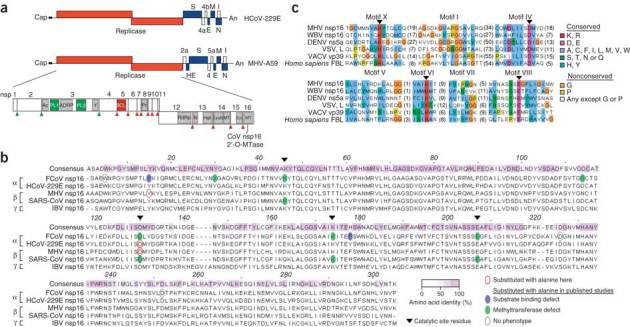
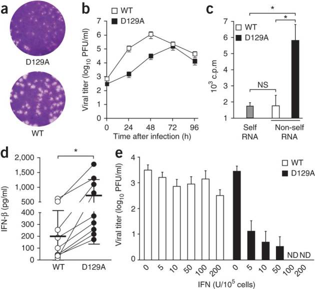
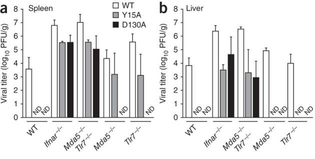
The 5' cap structures of higher eukaryote mRNAs have ribose 2'-O-methylation. Likewise, many viruses that replicate in the cytoplasm of eukaryotes have evolved 2'-O-methyltransferases to autonomously modify their mRNAs. However, a defined biological role for 2'-O-methylation of mRNA remains elusive. Here we show that 2'-O-methylation of viral mRNA was critically involved in subverting the induction of type I interferon. We demonstrate that human and mouse coronavirus mutants lacking 2'-O-methyltransferase activity induced higher expression of type I interferon and were highly sensitive to type I interferon. Notably, the induction of type I interferon by viruses deficient in 2'-O-methyltransferase was dependent on the cytoplasmic RNA sensor Mda5. This link between Mda5-mediated sensing of viral RNA and 2'-O-methylation of mRNA suggests that RNA modifications such as 2'-O-methylation provide a molecular signature for the discrimination of self and non-self mRNA.


Conflict of interest statement

The authors declare no competing financial interests.

Figures

Figure 1
Figure 1. Conservation of viral 2′-O-methyltransferases.
(a) Human and mouse coronavirus genomes, including viral open reading frames (boxes; labels above and below identify gene products). The replicase gene, including conserved domains and viral proteinase cleavage sites (upward arrowheads) that separate nsp1–nsp16, is enlarged. CoV nsp16 MTase, nsp16-associated 2′-O-methyltransferase. (b) ClustalW2 alignment of coronavirus nsp16 amino acid sequences, representative of α-, β- and γ-coronaviruses. Keys (bottom right) identify sequence conservation (amino acid identity), amino acid substitutions and phenotypes of mutant proteins. FCoV, feline coronavirus; MHV, MHV strain A59; SARS-CoV, severe acute respiratory syndrome coronavirus; IBV, infectious bronchitis virus. (c) Comparison of the sequences of viral and cellular methyltransferase motifs. WBV, white bream virus (order Nidovirales); DENV, dengue virus (family Flaviviridae); VSV, vesicular stomatitis virus (order Mononegavirales); VACV, vaccinia virus (family Poxviridae); FBL, fibrillarin (Homo sapiens). Right, key for amino acid similarity (single-letter codes) and conservation according to default ClustalX.
Figure 2
Figure 2. The HCoV 2′-O-methyltransferase mutant has altered replication kinetics and induction of and sensitivity to type I interferon.
(a) Plaque assay of HCoV-229E and HCoV-D129A. (b) Replication kinetics of wild-type HCoV-229E (WT) and the mutant HCoV-D129A (D129A) in MRC-5 cells infected at an MOI of 0.1, presented as viral titer in plaque-forming units (PFU). (c) Incorporation of 3H into poly(A)-containing RNA derived from mock-infected cells (Self RNA) or cells infected with HCoV-229E or HCoV-D129A (Non-self RNA) after in vitro 2′-O-methylation with VP39. NS, not significant (P > 0.05); *P < 0.01 (unpaired Student's t-test). (d) Enzyme-linked immunosorbent assay of IFN-β in supernatants of human blood–derived macrophages 24 h after infection with HCoV-229E or HCoV-D129A (MOI = 1). Each symbol represents an individual donor (n = 9); thick horizontal lines indicate the mean (thin horizontal lines, s.d.). *P < 0.005 (Wilcoxon matched-pairs test). (e) Plaque assay of viral titers in human blood–derived macrophages pretreated with increasing doses of IFN-α (horizontal axis) 4 h before infection with HCoV-229E or HCoV-D129A (MOI = 1), assessed 24 h after infection. ND, not detected. Data are representative of three experiments (a) or represent two (b,e) or three (c,d) independent experiments (average and s.e.m. of triplicates in b; mean and s.d. in c; error bars, s.d. of six samples in e).
Figure 3
Figure 3. MHV 2′-O-methyltransferase mutants induce IFN-β in an Mda5-dependent manner.
(a) Ethidium bromide staining (left) of poly(A)-containing RNA (300 ng) from cells infected with wild-type MHV-A59 (WT), MHV-Y15A (Y15A) or MHV-D130A (D130A), separated by electrophoresis through a 1% agarose gel; right margin (1–7), genomic and subgenomic mRNA; left margin, size in kilobases (kb). Right, incorporation of 3H into poly(A)-containing RNA from mock-infected cells or cells infected with MHV-A59, MHV-Y15A or MHV-D130A after in vitro 2′-O-methylation with VP39. (b) Replication kinetics of MHV-A59, MHV-Y15A and MHV-D130A in 17Cl1 cells after infection at an MOI of 1 or 0.0001. (ce) Enzyme-linked immunosorbent assay of IFN-β in supernatants of wild-type (WT; c), IFNAR-deficient (Ifnar−/−; d) or Mda5-deficient (Mda5−/−; e) macrophages (MΦ; 1 × 10⁶) infected with wild-type or mutant MHV or Sendai virus (SeV) at an MOI of 1 and assessed 15 h after infection. (f,g) Quantitative RT-PCR analysis of the kinetics of IFN-β mRNA expression in wild-type (f) or IFNAR-deficient (g) macrophages (1 × 10⁶) infected with MHV-A59, MHV-Y15A or MHV-D130A (MOI = 1), presented relative to its expression in uninfected cells. *P < 0.05, **P < 0.01 and ***P < 0.001 (unpaired Student's t-test). Data represent seven (a), two (b,f,g) or three (ce) independent experiments (mean and s.d. of seven (a) or six (cg) samples, or mean and s.e.m. of six samples (b)).
Figure 4
Figure 4. MHV 2′-O-methyltransferase mutants induce the nuclear localization of IRF3 in an Mda5-dependent manner.
(a) IRF3 in IFNAR-deficient or Mda5-deficient mouse macrophages infected with MHV-A59, MHV-Y15A or MHV-D130A at an MOI of 1 and stained at 3 h after infection for IRF3 (red) and with the DNA-intercalating dye DAPI (blue). Original magnification, ×20. Data are representative of three experiments. (b) Frequency of cells (infected as in a) with nuclear IRF3. *P < 0.01 and **P < 0.001 (unpaired Student's t-test). Data are representative of three experiments (mean and s.d. of five random fields with approximately 50–250 cells each).
Figure 5
Figure 5. Differences in the effect of type I interferon on the replication of MHV 2′-O-methyltransferase mutants.
(a,b) Kinetics of the replication of MHV-A59, MHV-Y15A or MHV-D130A in wild-type and Mda5-deficient mouse macrophages (1 × 10⁶) after infection at an MOI of 1 (a) or 0.0001 (b). Data represent two independent experiments (mean ± s.e.m. of five samples). (c,d) Titer of MHV-A59, MHV-Y15A or MHV-D130A at 24 h after infection (MOI = 1) of wild-type (c) or Mda5-deficient (d) macrophages (1 × 10⁵) pretreated with IFN-α (dose, horizontal axis) 4 h before infection. Data represent two independent experiments (mean and s.d. of four samples).
Figure 6
Figure 6. Restoration of MHV-D130A replication in IFIT1-deficient macrophages.
Kinetics of the replication of MHV-A59, MHV-Y15A or MHV-D130A after infection (MOI = 0.01) of wild-type mice (a) or IFIT1-deficient (b) macrophages (5 × 10⁵), assessed by plaque assay of viral titers in supernatants. Data represent two independent experiments (mean ± s.e.m. of four samples).
Figure 7
Figure 7. Deficiency in 2′-O-methyltransferase affects the recognition of virus by the innate immune system in vivo.
Viral titers in the spleen (a) and liver (b) of wild-type mice and mice deficient in IFNAR, Mda5 or TLR7 or both Mda5 and TLR7, assessed 48 h after infection with MHV-A59, MHV-Y15A or MHV-D130A (500 plaque-forming units injected intraperitoneally). Data represent two independent experiments (mean and s.d. of six samples).

Comment in

References

    1. Janeway CA., Jr. Approaching the asymptote? Evolution and revolution in immunology. Cold Spring Harb. Symp. Quant. Biol. 1989;54:1–13. doi: 10.1101/SQB.1989.054.01.003. - DOI - PubMed
    1. Takeuchi O, Akira S. Innate immunity to virus infection. Immunol. Rev. 2009;227:75–86. doi: 10.1111/j.1600-065X.2008.00737.x. - DOI - PMC - PubMed
    1. Loo YM, Gale M., Jr. Viral regulation and evasion of the host response. Curr. Top. Microbiol. Immunol. 2007;316:295–313. - PubMed
    1. Haller O, Weber F. Pathogenic viruses: smart manipulators of the interferon system. Curr. Top. Microbiol. Immunol. 2007;316:315–334. - PMC - PubMed
    1. Hui DJ, Terenzi F, Merrick WC, Sen GC. Mouse p56 blocks a distinct function of eukaryotic initiation factor 3 in translation initiation. J. Biol. Chem. 2005;280:3433–3440. doi: 10.1074/jbc.M406700200. - DOI - PubMed

Publication types

MeSH terms