Correlation of central corneal thickness and optic nerve head topography in patients with primary open-angle glaucoma
- PMID: 21217900
- PMCID: PMC3003855
- DOI: 10.4103/0974-620X.64231
Correlation of central corneal thickness and optic nerve head topography in patients with primary open-angle glaucoma
Abstract
Purpose: To evaluate whether changes in optic nerve head topography and visual field in patients with primary open-angle (POAG) are related to central corneal thickness (CCT).
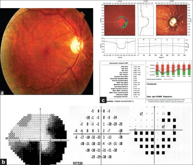
Materials and methods: Eighty eyes of 50 patients with POAG underwent ophthalmic examination; optic nerve head imaging with the Heidelberg Retina Tomography II (HRT II), ultrasound corneal pachymetry, and visual field evaluation with the Humphrey visual field analyser (program 24-2). Correlation between CCT, age, gender, family history of glaucoma, visual acuity, intraocular pressure (IOP), optic disc surface area, vertical and horizontal cup: disc ratios, neuroretinal rim area, mean deviation of visual field, and number of glaucoma medications was analyzed. Patients were divided into a thin CCT group <540 μm or a thick CCT group >540 μm. Pearson correlation was used for correlation coefficient and a P value of <0.05 was considered statistically significant.
Results: THIN CCT WAS SIGNIFICANTLY CORRELATED WITH VERTICAL AND HORIZONTAL CUP: disc ratios, neuroretinal rim area loss, and smaller optic disc surface area (r=0.043, r=0.021, r=0.036, and 0.031 respectively). Thin CCT was also significantly associated with worsened mean deviation of visual field, and increased number of glaucoma medications (r=0.065 and r=0.423). Patients with positive family history correlated with with greater vertical cup: disc ratio, and more glaucoma medications but this was not statistically significant.
Conclusions: In patients with POAG those with thinner CCT are likely to develop greater glaucomatous optic nerve and visual field damages than those with a thicker CCT.
Keywords: Central corneal thickness; optic nerve head; primary open angle glaucoma; visual field.
Conflict of interest statement
Figures
Similar articles
-
Comparison of optic nerve head topography and visual field in eyes with open-angle and angle-closure glaucoma.Ophthalmology. 2008 Feb;115(2):239-245.e2. doi: 10.1016/j.ophtha.2007.03.086. Ophthalmology. 2008. PMID: 18082888
-
Relationships between central corneal thickness and optic disc topography in eyes with glaucoma, suspicion of glaucoma, or ocular hypertension.Clin Ophthalmol. 2008 Sep;2(3):591-9. doi: 10.2147/opth.s2814. Clin Ophthalmol. 2008. PMID: 19668759 Free PMC article.
-
The relationship between central corneal thickness and optic disc size in patients with primary open-angle glaucoma in a hospital-based population.Acta Ophthalmol. 2011 Sep;89(6):556-9. doi: 10.1111/j.1755-3768.2009.01746.x. Epub 2009 Oct 30. Acta Ophthalmol. 2011. PMID: 19878114
-
Central corneal thickness and correlation to optic disc size: a potential link for susceptibility to glaucoma.Br J Ophthalmol. 2007 Jan;91(1):26-8. doi: 10.1136/bjo.2006.106039. Epub 2006 Sep 14. Br J Ophthalmol. 2007. PMID: 16973656 Free PMC article.
-
Central Corneal Thickness and Glaucoma Risk: The Importance of Corneal Pachymetry in Screening Adults Over 50 and Glaucoma Suspects.Clin Ophthalmol. 2025 Feb 15;19:563-570. doi: 10.2147/OPTH.S498422. eCollection 2025. Clin Ophthalmol. 2025. PMID: 39974660 Free PMC article. Review.
Cited by
-
Measurement of Central Corneal Thickness Using Ultrasound Pachymetry and Orbscan II in Normal Eyes.J Ophthalmic Vis Res. 2015 Jan-Mar;10(1):4-9. doi: 10.4103/2008-322X.156084. J Ophthalmic Vis Res. 2015. PMID: 26005545 Free PMC article.
-
Corneal Segmentation Analysis Increases Glaucoma Diagnostic Ability of Optic Nerve Head Examination, Heidelberg Retina Tomograph's Moorfield's Regression Analysis, and Glaucoma Probability Score.J Ophthalmol. 2015;2015:215951. doi: 10.1155/2015/215951. Epub 2015 May 27. J Ophthalmol. 2015. PMID: 26180641 Free PMC article.
-
Integrating neuroprotection, antioxidative effects, and precision medicine in glaucoma management with bioactive compounds.Biomed Pharmacother. 2025 Sep;190:118319. doi: 10.1016/j.biopha.2025.118319. Epub 2025 Jul 18. Biomed Pharmacother. 2025. PMID: 40683209 Free PMC article. Review.
-
Comparison of central corneal thickness using non-contact tono-pachymeter (Tonopachy) with ultrasound pachymetry in normal children and in children with refractive error.Indian J Ophthalmol. 2021 Aug;69(8):2053-2059. doi: 10.4103/ijo.IJO_364_21. Indian J Ophthalmol. 2021. PMID: 34304177 Free PMC article.
-
Major review: Molecular genetics of primary open-angle glaucoma.Exp Eye Res. 2017 Jul;160:62-84. doi: 10.1016/j.exer.2017.05.002. Epub 2017 May 10. Exp Eye Res. 2017. PMID: 28499933 Free PMC article. Review.
References
-
- Herndon LW, Weizer JS, Stinnett SS. Central corneal thickness as a risk factor for advanced glaucoma damage. Arch Ophthalmol. 2004;122:17–21. - PubMed
-
- Shih CY, Graff Zivin JS, Trokel SL, Tsai JC. Clinical significance of central corneal thickness in the management of glaucoma. Arch Ophthalmol. 2004;122:1270–5. - PubMed
-
- Muir KW, Jin J, Freedman SF. Central corneal thickness and its relationship to intraocular pressure in children. Ophthalmology. 2004;111:2220–3. - PubMed
-
- Kohlhaas M, Boehm AG, Spoerl E, Pürsten A, Grein HJ, Pillunat LE. Effect of central corneal thickness, corneal curvature, and axial length on applanation tonometry. Arch Ophthalmol. 2006;124:471–6. - PubMed
-
- Gordon MO, Beiser JA, Brandt JD, Heuer DK, Higginbotham EJ, Johnson CA, et al. The Ocular Hypertension Treatment Study: baseline factors that predict the onset of primary open angle glaucoma. Arch Ophthalmol. 2002;120:714–20. - PubMed