

Detail microscopic analysis of deep fascia of lower limb and its surgical implication
- PMID: 21217969
- PMCID: PMC3010771
- DOI: 10.4103/0970-0358.73424
Detail microscopic analysis of deep fascia of lower limb and its surgical implication
Abstract
Background: The knowledge regarding the structural details of deep fascia remains inadequate. It was described to be relatively avascular having predominantly protective function. Anatomical and surgical studies revealed that it had associated vascular arcade and hence incorporated it to ascertain additional vascularity to the flaps. However, not much importance has been directed towards the detailed study of the various constituents of deep fascia in order to explain its physiological and clinical implications. Therefore, this study was undertaken to unveil these details.
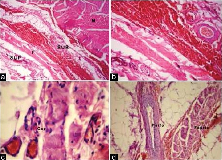
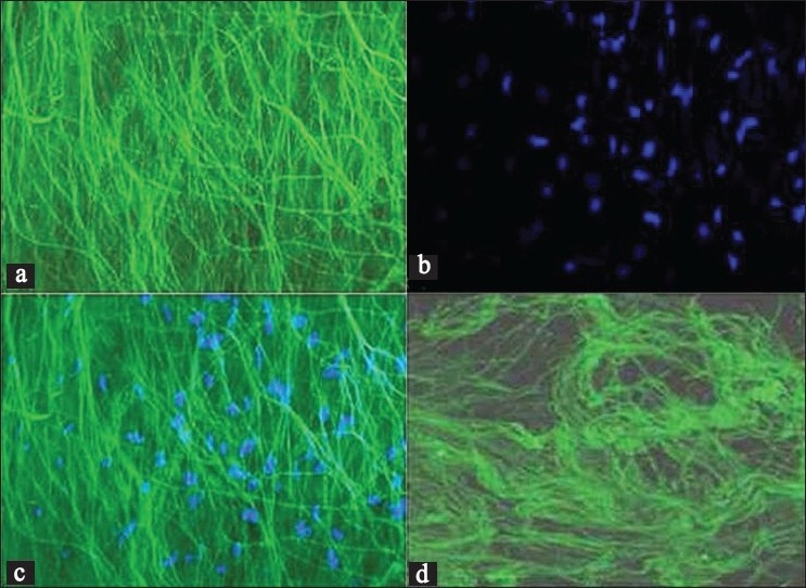
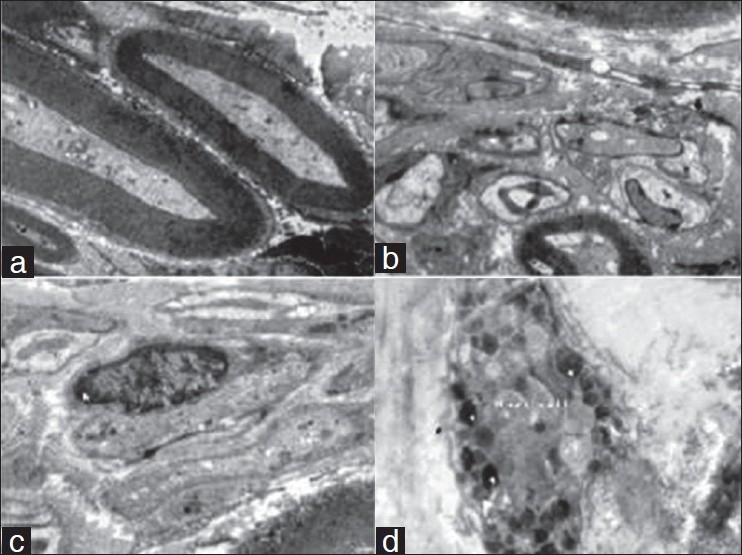
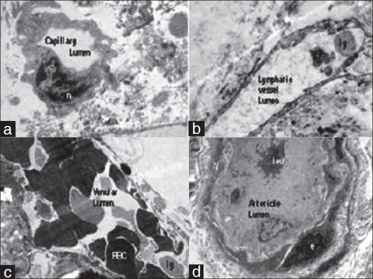
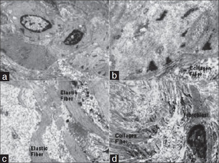
Materials and methods: Fifty fresh specimens of human deep fascia overlying the gastrocnemius muscle were analyzed regarding the (i) vasculature, (ii) matrix, and (iii) other structural elements. The deep fascia was procured in three forms; (a) both the layers, (b) superficial layer, and (c) deep layer. Detail study was conducted by light, confocal, and electron microscopy.
Results: Under light microscopy, blood vessels including capillaries were seen associated with both the layers. Perforators traversing the intra-fascial plane could be visualized. Confocal microscope optical sections showed well-organized bright fluorescent collagen fibers and nuclei of various cells. Electron microscopic evaluation revealed many interesting constituents which are relatively unknown to the anatomist and clinicians. There were arterioles, capillaries, venules, lymphatics, nerves, mast cells, and myofibroblasts apart from collagen and elastic fibers.
Conclusion: The detail structural analysis of deep fascia provided the clue to its rich vascularity and other structural constituents. They all contribute to enhance the vascularity and maintenance of the physiological functions of fasciocutaneous, adipofascial, and fascial flaps, frequently used for reconstructions. Thus, incorporation of deep fascia in the flaps during reconstruction is highly beneficial for ensuring optimal vascularity.
Keywords: Confocal microscopy; deep fascia; electron microscopy; flap; microscopic anatomy.
Conflict of interest statement
Figures
References
-
- Geneser F. Textbook of histology. Copenhagen: Munksgaard Lea and Febiger; 1986.
-
- Standring S, Ellis H, Healy J, Johnson D, Williams A. Gray’s anatomy. 39th ed. London: Churchill Livingstone; 2005.
-
- Ponten B. The fasciocutaneous flap: Its use in soft tissue defects of the lower leg. Br J Plast Surg. 1981;34:215–20. - PubMed
-
- Donski PK, Fogdestam I. Distally based fasciocutaneous flap from the sural region. Scand J Plast Reconstr Surg. 1983;17:191–6. - PubMed
-
- Carriquiry C, Aparecida Costa M, Vascomz LO. An anatomic study of the septocutaneous vessels of the leg. Plast Reconstr Surg. 1985;76:354–60. - PubMed
LinkOut - more resources
Full Text Sources