

Contrast-enhanced endoscopic ultrasonography
- PMID: 21218082
- PMCID: PMC3016678
- DOI: 10.3748/wjg.v17.i1.42
Contrast-enhanced endoscopic ultrasonography
Abstract
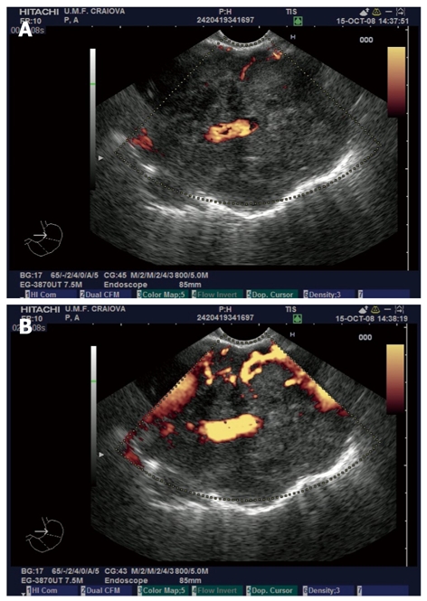
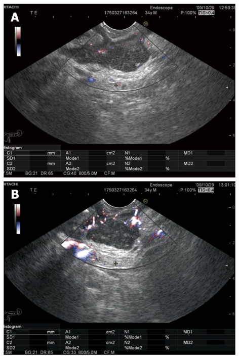
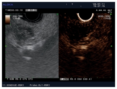
Contrast agents are increasingly being used to characterize the vasculature in an organ of interest, to better delineate benign from malignant pathology and to aid in staging and directing therapeutic procedures. We review the mechanisms of action of first, second and third generation contrast agents and their use in various endoscopic procedures in the gastrointestinal tract. Various applications of contrast-enhanced endoscopic ultrasonography include differentiating benign from malignant mediastinal lymphadenopathy, assessment of depth of invasion of esophageal, gastric and gall bladder cancers and visualization of the portal venous system and esophageal varices. In addition, contrast agents can be used to differentiate pancreatic lesions. The use of color Doppler further increases the ability to diagnose and differentiate various pancreatic malignancies. The sensitivity of power Doppler sonography to depict tumor neovascularization can be increased by contrast agents. Contrast-enhanced harmonic imaging is a useful aid in identifying the tumor vasculature and studying pancreatic microperfusion. In the future, these techniques could potentially be used to quantify tumor perfusion, to assess and monitor the efficacy of antiangiogenic agents, to assist targeted drug delivery and allow molecular imaging.
Keywords: Contrast media; Doppler ultrasonography; Endoscopic ultrasonography; Gastrointestinal neoplasms; Pancreatic cancer.
Figures
References
-
- Keller MW, Feinstein SB, Watson DD. Successful left ventricular opacification following peripheral venous injection of sonicated contrast agent: an experimental evaluation. Am Heart J. 1987;114:570–575. - PubMed
-
- Dietrich CF, Ignee A, Braden B, Barreiros AP, Ott M, Hocke M. Improved differentiation of pancreatic tumors using contrast-enhanced endoscopic ultrasound. Clin Gastroenterol Hepatol. 2008;6:590–597.e1. - PubMed
-
- Goldberg BB, Hilpert PL, Burns PN, Liu JB, Newman LM, Merton DA, Witlin LA. Hepatic tumors: signal enhancement at Doppler US after intravenous injection of a contrast agent. Radiology. 1990;177:713–717. - PubMed
-
- Hirooka Y, Goto H, Ito A, Hayakawa S, Watanabe Y, Ishiguro Y, Kojima S, Hayakawa T, Naitoh Y. Contrast-enhanced endoscopic ultrasonography in pancreatic diseases: a preliminary study. Am J Gastroenterol. 1998;93:632–635. - PubMed
-
- Hirooka Y, Naitoh Y, Goto H, Ito A, Hayakawa S, Watanabe Y, Ishiguro Y, Kojima S, Hashimoto S, Hayakawa T. Contrast-enhanced endoscopic ultrasonography in gallbladder diseases. Gastrointest Endosc. 1998;48:406–410. - PubMed
Publication types
MeSH terms
Substances
LinkOut - more resources
Full Text Sources
Other Literature Sources
