

New endoscopic approaches in IBD
- PMID: 21218085
- PMCID: PMC3016681
- DOI: 10.3748/wjg.v17.i1.63
New endoscopic approaches in IBD
Abstract
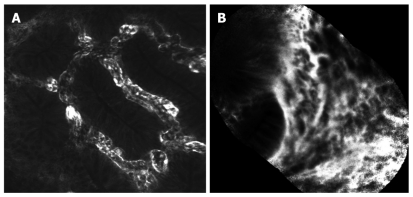
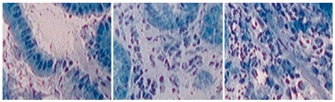
Recent advances in endoscopic imaging techniques have revolutionized the diagnostic approach of patients with inflammatory bowel disease (IBD). New, emerging endoscopic imaging techniques visualized a plethora of new mucosal details even at the cellular and subcellular level. This review offers an overview about new endoscopic techniques, including chromoendoscopy, magnification endoscopy, spectroscopy, confocal laser endomicroscopy and endocytoscopy in the face of IBD.
Keywords: Chromoendoscopy; Crohn’s disease; Endocytoscopy; Endomicroscopy; Endoscopy; Fluorescence endoscopy; Fujinon intelligent color enhancement; Inflammatory bowel disease; Narrow band imaging; Spectroscopy; Ulcerative colitis; i-Scan.
Figures
Similar articles
-
New endoscopic imaging techniques in surveillance of inflammatory bowel disease.World J Gastrointest Endosc. 2015 Mar 16;7(3):230-6. doi: 10.4253/wjge.v7.i3.230. World J Gastrointest Endosc. 2015. PMID: 25789093 Free PMC article. Review.
-
Cancer risk in IBD: how to diagnose and how to manage DALM and ALM.World J Gastroenterol. 2011 Jul 21;17(27):3184-91. doi: 10.3748/wjg.v17.i27.3184. World J Gastroenterol. 2011. PMID: 21912466 Free PMC article. Review.
-
Advanced Endoscopic Imaging Techniques for the Study of Colonic Mucosa in Patients with Inflammatory Bowel Disease.Rom J Intern Med. 2016 Jan-Mar;54(1):11-23. doi: 10.1515/rjim-2015-0050. Rom J Intern Med. 2016. PMID: 27141566 Review.
-
Advanced endoscopic imaging techniques in Crohn's disease.J Crohns Colitis. 2014 Apr;8(4):261-9. doi: 10.1016/j.crohns.2013.09.004. Epub 2013 Sep 29. J Crohns Colitis. 2014. PMID: 24080247 Review.
-
Confocal Laser Endomicroscopy in the Evaluation of Inflammatory Bowel Disease.Inflamm Bowel Dis. 2019 Jul 17;25(8):1302-1312. doi: 10.1093/ibd/izz021. Inflamm Bowel Dis. 2019. PMID: 30877772 Review.
Cited by
-
Advanced endoscopic imaging for diagnosis of Crohn's disease.Gastroenterol Res Pract. 2012;2012:301541. doi: 10.1155/2012/301541. Epub 2011 Nov 24. Gastroenterol Res Pract. 2012. PMID: 22144998 Free PMC article.
-
Rediscovering histology: what is new in endoscopy for inflammatory bowel disease?Therap Adv Gastroenterol. 2021 Apr 16;14:17562848211005692. doi: 10.1177/17562848211005692. eCollection 2021. Therap Adv Gastroenterol. 2021. PMID: 33948114 Free PMC article. Review.
-
Role of Advanced Endoscopic Imaging Techniques in the Management of Inflammatory Bowel Disease.Clin Endosc. 2017 Sep;50(5):424-428. doi: 10.5946/ce.2017.143. Epub 2017 Sep 29. Clin Endosc. 2017. PMID: 29017290 Free PMC article. Review.
-
Current status of image-enhanced endoscopy in inflammatory bowel disease.Clin Endosc. 2023 Sep;56(5):563-577. doi: 10.5946/ce.2023.070. Epub 2023 Sep 26. Clin Endosc. 2023. PMID: 37793436 Free PMC article. Review.
-
New endoscopic imaging techniques in surveillance of inflammatory bowel disease.World J Gastrointest Endosc. 2015 Mar 16;7(3):230-6. doi: 10.4253/wjge.v7.i3.230. World J Gastrointest Endosc. 2015. PMID: 25789093 Free PMC article. Review.
References
-
- Connell WR, Lennard-Jones JE, Williams CB, Talbot IC, Price AB, Wilkinson KH. Factors affecting the outcome of endoscopic surveillance for cancer in ulcerative colitis. Gastroenterology. 1994;107:934–944. - PubMed
-
- Kiesslich R, Fritsch J, Holtmann M, Koehler HH, Stolte M, Kanzler S, Nafe B, Jung M, Galle PR, Neurath MF. Methylene blue-aided chromoendoscopy for the detection of intraepithelial neoplasia and colon cancer in ulcerative colitis. Gastroenterology. 2003;124:880–888. - PubMed
-
- Hurlstone DP, McAlindon ME, Sanders DS, Koegh R, Lobo AJ, Cross SS. Further validation of high-magnification chromoscopic-colonoscopy for the detection of intraepithelial neoplasia and colon cancer in ulcerative colitis. Gastroenterology. 2004;126:376–378. - PubMed
-
- Matsumoto T, Kudo T, Jo Y, Esaki M, Yao T, Iida M. Magnifying colonoscopy with narrow band imaging system for the diagnosis of dysplasia in ulcerative colitis: a pilot study. Gastrointest Endosc. 2007;66:957–965. - PubMed
-
- van den Broek FJ, Fockens P, van Eeden S, Reitsma JB, Hardwick JC, Stokkers PC, Dekker E. Endoscopic tri-modal imaging for surveillance in ulcerative colitis: randomised comparison of high-resolution endoscopy and autofluorescence imaging for neoplasia detection; and evaluation of narrow-band imaging for classification of lesions. Gut. 2008;57:1083–1089. - PMC - PubMed
Publication types
MeSH terms
LinkOut - more resources
Full Text Sources
Medical
