. 2010 Jan 1;250(1):1-5.
doi: 10.1088/1742-6596/250/1/012007.

Fast, large field-of-view, telecentric optical-CT scanning system for 3D radiochromic dosimetry

Affiliations

Fast, large field-of-view, telecentric optical-CT scanning system for 3D radiochromic dosimetry

A Thomas et al. J Phys Conf Ser. .

Abstract

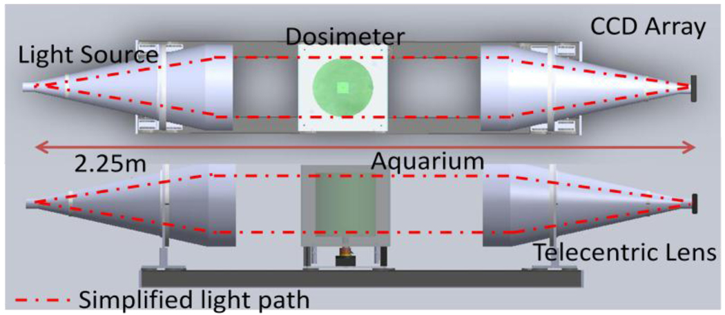
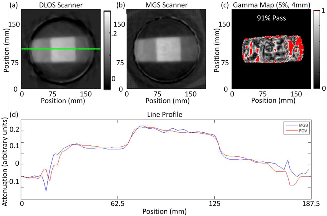
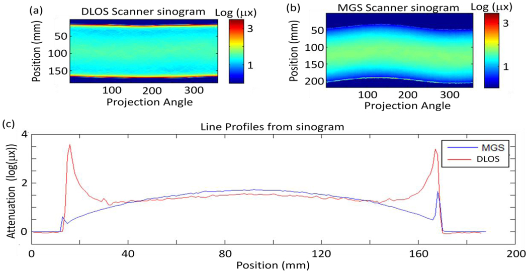
We describe initial experiences with an in-house, fast, large field-of-view optical-CT telecentric scanner (the Duke Large field of view Optical-CT Scanner (DLOS)). The DLOS system is designed to enable telecentric optical-CT imaging of dosimeters up to 24 cm in diameter with a spatial resolution of 1 mm(3), in approximately 10 minutes. These capabilities render the DLOS system a unique device at present. The system is a scaled up version of early prototypes in our lab. This scaling introduces several challenges, including the accurate measurement of a greatly increased range of light attenuation within the dosimeter, and the need to reduce even minor reflections and scattered light within the imaging chain. We present several corrections and techniques that enable accurate, low noise, 3D dosimetery with the DLOS system.


Figures

Figure 1
Figure 1
A schematic of the DLOS system in a top and side view showing the collimated LED light source, acquarium, rotation stage, telecentric lens and detector array.
Figure 2
Figure 2
series of images showing the internal scatter present within the lens. (a) Image acquired with the lens cover on, (b) an image acquired without the lens cover and (c) an image captured with the lens cover in position to let a “sliver” of light through. Notice the contrast between (a) & (b).
Figure 3
Figure 3
(a) Image of a filter wheel with known optical densities ranging from 1 – 4 converted to OD. The OD values of ND = 1 & 2 match the known filter values, however, the higher values, ND = 3 & 4 are not near their known values. (b) An image of the same filter wheel taken with a series of 3 images with increasing exposure times to create an accurate (within 4%), high dynamic range image. This represents an image accurately covering 80 dB of dynamic range.
Figure 4
Figure 4
Sketch of the two field 6 cm × 6 cm plan delivered to the 15 cm × 12 cm dosimeter.
Figure 5
Figure 5
(a) Reconstructed slice from the DLOS system. (b) The same reconstructed slice from the MGS scanner. Both scans were acquired with 360 projections over 360 degrees. (c) A 5%, 4 mm, 5% threshold for the slices shown in (a) and (b) with a 91% pass rate calculated only from pixels within the dosimeter. (d) Line profiles taken from the green line in slices (a) and (b).
Figure 6
Figure 6
(a) Flood corrected sinogram from prescan captured with the large DLOS system. (b) Flood corrected sinogram from the prescan acquired with the MGS scanner & (c) a set of line profiles from each sinogram in (a) and (b) showing the differences in the scanners. Most evident are the edge artefacts present in each scanner, however, those in the DLOS system are much more pronounced and alters the shape of the curve for the 1st and last 25 mm of each projection angle. The same fluid was used in each scan and shows how the MGS scanner is much less sensitive to the refractive index match.

References

    1. Doran SJ, et al. A CCD-based optical CT scanner for high-resolution 3D imaging of radiation dose distributions: equipment specifications, optical simulations and preliminary results. Phys Med Biol. 2001;46(12):3191–3213. - PubMed
    1. Oldham M, et al. High resolution gel-dosimetry by optical-CT and MR scanning. Med Phys. 2001;28(7):1436–1445. - PubMed
    1. Sakhalkar HS, Oldham M. Fast, high-resolution 3D dosimetry utilizing a novel optical-CT scanner incorporating tertiary telecentric collimation. Med Phys. 2008;35(1):101–111. - PMC - PubMed
    1. Wolodzko JG, Marsden C, Appleby A. CCD imaging for optical tomography of gel radiation dosimeters. Med Phys. 1999;26(11):2508–2513. - PubMed
    1. Clift C, et al. Toward acquiring comprehensive radiosurgery field commissioning data using the PRESAGE/optical-CT 3D dosimetry system. Phys Med Biol. 55(5):1279–1293. - PMC - PubMed