Review
. 2011:62:483-506.
doi: 10.1146/annurev.physchem.012809.103450.

Thermodynamics and mechanics of membrane curvature generation and sensing by proteins and lipids

Affiliations
Review

Thermodynamics and mechanics of membrane curvature generation and sensing by proteins and lipids

Tobias Baumgart et al. Annu Rev Phys Chem. 2011.

Abstract

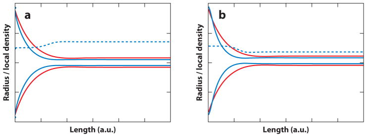
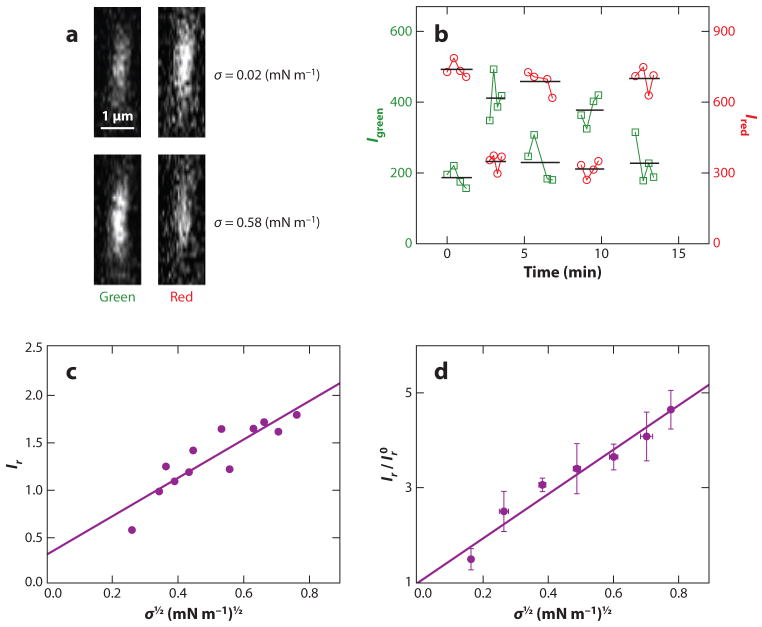
Research investigating lipid membrane curvature generation and sensing is a rapidly developing frontier in membrane physical chemistry and biophysics. The fast recent progress is based on the discovery of a plethora of proteins involved in coupling membrane shape to cellular membrane function, the design of new quantitative experimental techniques to study aspects of membrane curvature, and the development of analytical theories and simulation techniques that allow a mechanistic interpretation of quantitative measurements. The present review first provides an overview of important classes of membrane proteins for which function is coupled to membrane curvature. We then survey several mechanisms that are assumed to underlie membrane curvature sensing and generation. Finally, we discuss relatively simple thermodynamic/mechanical models that allow quantitative interpretation of experimental observations.


Figures

Figure 1
Figure 1
Confocal micrographs demonstrating protein curvature partitioning among tubular lipid membranes with high membrane curvature, and giant unilamellar vesicle (GUV) membranes with negligible curvature. The experimental setup is as referred to in the text (see and for further description). Briefly, a bead (present on the left in each micrograph) with controllable position was specifically adhered to a GUV (visible on the right of each micrograph) and laterally translated to form a tubular membrane connected to the GUV, except in panel g (41). Unless otherwise indicated, GUVs were composed of DOPC with auxiliary lipids mixed at the mole fractions specified, additionally contained Texas Red-DHPE and Biotin-PEG2000-DSPE, and were formed by electroswelling in sucrose solution equi-osmolar to the respective experimental solution conditions. Unless otherwise indicated, each protein shown was conjugated to AlexaFluor 488. The respective references reporting tubulation by these proteins in vitro are included, except for cholera toxin subunit B (CTB), which provides a comparative example. Scale bars (yellow lines) are equal to 3 μm in each micrograph. (a) Drosophila amphiphysin N-BAR (DA N-BAR) (9): 60-nM protein, GUV with 20% DOPS and 12% cholesterol (σ = 0.59 mN m−1), in 1-M sucrose, 15-mM NaCl, and HEPES pH 7.4 (unpublished data from our lab). (b) Rat endophilin A1 N-BAR (6): 150-nM protein, GUV with 25% DOPG (σ = 0.22 mN m−1), in 33-mM NaCl, and HEPES pH 7.4 (unpublished data from our lab). (c) Rgd1 F-BAR [of the SrGAP/Gas7 subfamily (24); see 130]: 190-nM protein, GUV with 40% DOPG (σ = 0.10 mN m−1), in 130-mM NaCl, and HEPES pH 7.4 (unpublished data from our lab). (d) Dynamin (4): conditions as in Reference . Images adapted from Reference with permission. (e) Epsin1 ENTH-GFP (8): conditions as in Reference (σ = 0.31 mN m−1) (unpublished data from our lab). (f) Sar1 (13, 132): 1-μM protein (H79G), GUV with 5% DOPS and 10% cholesterol (σ = 0.09 mN m−1), in 2-mM GTP, 9-mM KOAc, 2.5-mM Mg (OAc)2, 2.5-mM EDTA, and HEPES pH 7.2 (unpublished data from our lab). (g) ArfGAP1: conditions and tubule formation as described in Reference . Images adapted from Reference by permission from Macmillan Publishers Ltd: The EMBO Journal, copyright 2010. (h) α-synuclein (22): 0.5-μM protein, GUV with 50% DOPG (σ = 0.36 mN m−1), in PBS (unpublished data from our lab). (i) CTB: conditions as in Reference (σ = 0.04 mN m−1). Images adapted from Reference , copyright 2009, with permission from Elsevier.
Figure 2
Figure 2
Mechanisms of membrane curvature generation and sensing.
Figure 3
Figure 3
Curvature sensing of fluorescence labeled lipid-like amphiphiles as revealed by (a) the single liposome curvature (SLiC) assay (35) and (b) the tube/giant unilamellar vesicle (GUV) assay (unpublished data from our lab). The comparison employs identical curvature-partitioning molecules [fluorescein-C12 (C12 or FL-C12) and TR-DHPE]. The SLiC assay is described in detail in Reference , and the tube/GUV assay is described in Reference . Whereas the SLiC assay suggests effective curvature sensing by lipids, the tube/GUV assay does not reveal measurable curvature sorting over the indicated range of tube radii. Panel a adapted with permission from Macmillan Publishers Ltd: Nature Chemical Biology, copyright 2009.
Figure 4
Figure 4
Schematic comparison of (a) the tube/giant unilamellar vesicle (GUV) assay and (b) the single liposome curvature (SLiC) assay. Unique features of the tube/GUV assay are the possibility of rapid equilibration of lipid monolayer density changes associated with tube-curvature changes by membrane area exchange with the large (GUV) membrane reservoir. Furthermore, in the GUV/tube assay the rapid exchange (38) of curvature-partitioning molecules among the tube monolayer and connected GUV monolayer allows rapid thermodynamic equilibration after curvature changes. It is therefore currently unclear to what extent lipid flipping may perturb the interpretation of lipid partitioning data obtained from the tube/GUV assay. Unique to the SLiC assay is that curvature-sensing molecules are added from an aqueous solution to vesicles of different curvatures. It is currently unclear to what extent outer and inner leaflets of vesicles equilibrate during SLiC experiments, and it is unclear to what extent lipid exchange among membranes of different vesicles is a process fast enough to allow for thermodynamic equilibration necessary for analysis with thermodynamic equilibrium models.
Figure 5
Figure 5
Computed membrane shapes obtained from the numerical integration of differential shape equations assuming boundary conditions that lead to an approximate catenoid shape with low curvature (left in both panels) and a tubular membrane (right in both panels). The shapes in both panels were obtained using identical parameters and boundary conditions (total membrane area A = 4π, length of the membrane shape L = 3, and lateral tension σ = 20). (a) Membrane shape (solid blue line) calculated assuming a curvature-composition coupling coefficient of Λ = −0.5, implying positive spontaneous curvature of a curvature-partitioning molecule added at a mole fraction of ϕ = 0.5 in a binary mixture additionally containing a species with negligible spontaneous curvature. Positive molecular spontaneous curvature leads to the enrichment of curvature-partitioning molecules in the outer leaflet of the tubular membrane (dashed blue line). For comparison, a shape has been added that was computed assuming Λ = 0 (solid red line); for vanishing curvature-composition coupling, a larger tube radius is observed. (b) Identical conditions as in panel a except that Λ = 0.5 was assumed, implying negative molecular spontaneous curvature and depletion (dashed blue line) of the curvature-partitioning molecule from the tubular membrane. In this case as well, a thinner tube (blue solid line) is observed compared to a membrane in the absence of curvature partitioning (red solid line).
Figure 6
Figure 6
Quantitative experimental measurements of curvature partitioning based on the tube/giant unilamellar vesicle assay, as demonstrated with the example of ENTH-GFP. (a) Representative tube fluorescence cross-section images comparing ENTH-GFP (green channel of the confocal fluorescence microscope) and lipid dye (red channel) at two different lateral tensions. Whereas ENTH-GFP fluorescence increases with an increase of lateral tension (coupled with an increase in membrane curvature), the lipid fluorescence intensity is observed to decrease with increasing tension as expected (38). (b) Demonstration of reversible fluorescence intensity changes under cycling membrane tension, demonstrating thermodynamic equilibrium conditions necessary for thermodynamic analysis. (c) Relative fluorescence intensity values Ir (expressed as protein versus lipid fluorescence) as a function of applied membrane tension of a tubular membrane. An approximately linear relationship is obtained. (d) The intercept resulting from extrapolation to zero tension is used to normalize multiple data sets. The slope of this graph can be related to the curvature-composition coupling coefficient Λ (42). For further details, we refer readers to Reference . Adapted with permission from Reference . Copyright 2010 American Chemical Society.

References

    1. Zimmerberg J, Kozlov MM. How proteins produce cellular membrane curvature. Nat Rev Mol Cell Biol. 2006;7:9–19. - PubMed
    1. McMahon HT, Gallop JL. Membrane curvature and mechanisms of dynamic cell membrane remodelling. Nature. 2005;438:590–96. - PubMed
    1. Frost A, Unger VM, De Camilli P. The BAR domain superfamily: membrane-molding macromolecules. Cell. 2009;137:191–96. - PMC - PubMed
    1. Sweitzer SM, Hinshaw JE. Dynamin undergoes a GTP-dependent conformational change causing vesiculation. Cell. 1998;93:1021–29. - PubMed
    1. Takei K, Slepnev VI, Haucke V, De Camilli P. Functional partnership between amphiphysin and dynamin in clathrin-mediated endocytosis. Nat Cell Biol. 1999;1:33–39. - PubMed

Publication types

LinkOut - more resources