Review
. 2011:29:273-93.
doi: 10.1146/annurev-immunol-031210-101317.

Immunoglobulin responses at the mucosal interface

Affiliations
Review

Immunoglobulin responses at the mucosal interface

Andrea Cerutti et al. Annu Rev Immunol. 2011.

Abstract

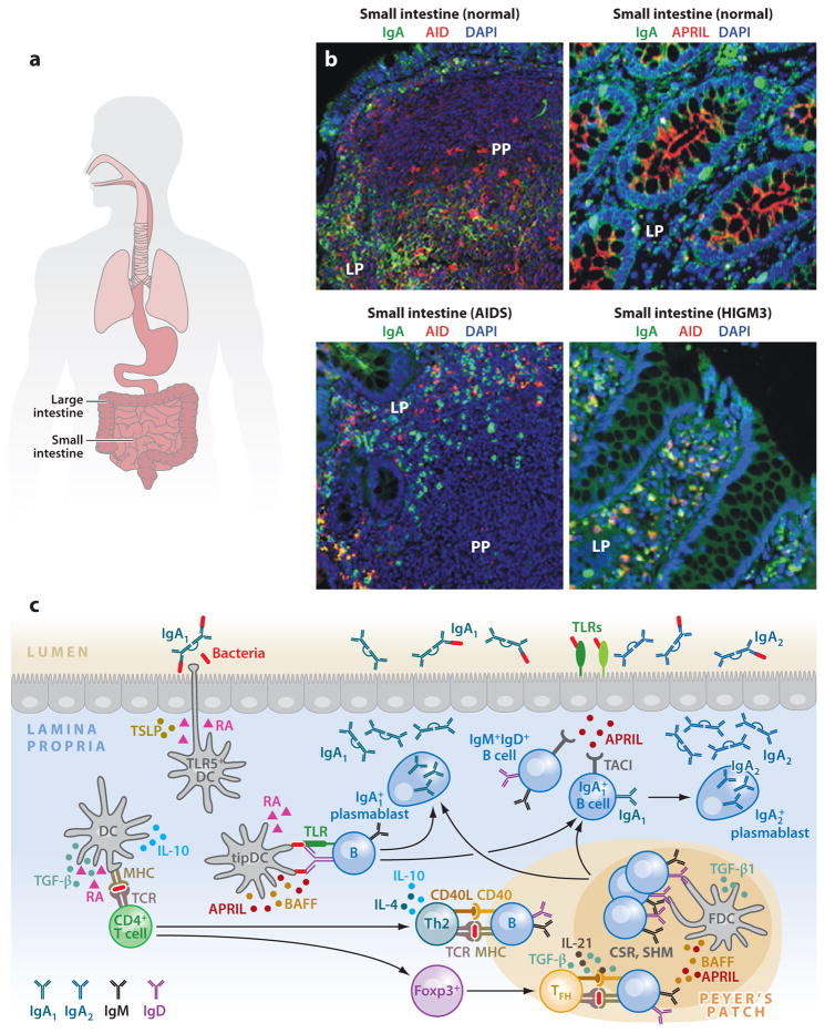
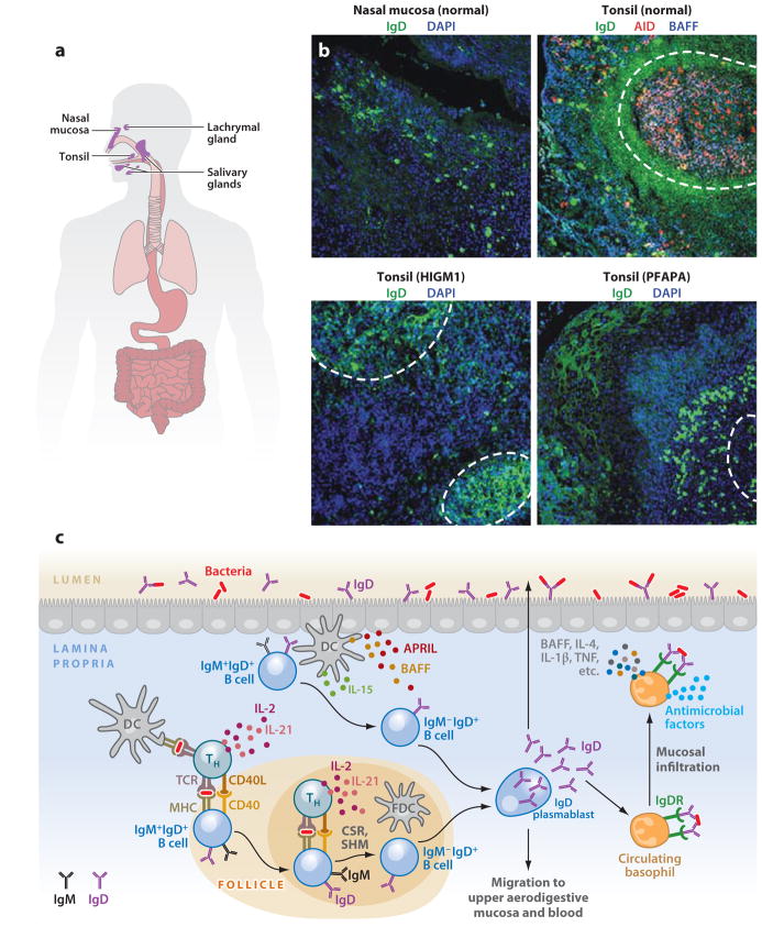
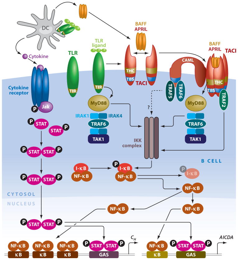
Mucosal surfaces are colonized by large communities of commensal bacteria and represent the primary site of entry for pathogenic agents. To prevent microbial intrusion, mucosal B cells release large amounts of immunoglobulin (Ig) molecules through multiple follicular and extrafollicular pathways. IgA is the most abundant antibody isotype in mucosal secretions and owes its success in frontline immunity to its ability to undergo transcytosis across epithelial cells. In addition to translocating IgA onto the mucosal surface, epithelial cells educate the mucosal immune system as to the composition of the local microbiota and instruct B cells to initiate IgA responses that generate immune protection while preserving immune homeostasis. Here we review recent advances in our understanding of the cellular interactions and signaling pathways governing IgA production at mucosal surfaces and discuss new findings on the regulation and function of mucosal IgD, the most enigmatic isotype of our mucosal antibody repertoire.


Figures

Figure 1
Figure 1
IgA responses in the intestinal mucosa. (a) Scheme of human MALT, including intestinal mucosa. (b) Immunofluorescence analysis of gut mucosa from healthy, HIGM3, and AIDS donors stained for IgA ( green), AID or APRIL (red ), and nuclei (DAPI staining, blue). Left panels show Peyer’s patches (PPs) and lamina propria (LP); right panels show only LP. Original magnification, ×20. (c) Scheme of mucosal IgA responses. Antigen-sampling DCs receive conditioning signals from TLR-activated intestinal epithelial cells (IECs) via thymic stromal lymphopoietin (TSLP) and retinoic acid (RA). Various DC subsets releasing TGF-β, IL-10, RA, and nitric oxide initiate IgA responses in PPs by inducing Th2, Treg, and Treg-derived T follicular helper (TFH) cells that activate follicular B cells via CD40L, TGF-β, IL-4, IL-10, and IL-21. In addition, DCs activate some B cells in the LP via BAFF, APRIL, and RA. These molecules are also used by TLR-activated IECs to induce local IgA production, including sequential switching from IgA1 to IgA2, as well as plasma cell differentiation and survival. (Additional abbreviations used in figure: AID, activation-induced cytidine deaminase; APRIL, a proliferation-inducing ligand; BAFF, B cell–activating factor; CSR, class switch recombination; DC, dendritic cell; HIGM3, hyper-IgM 3; MALT, mucosa-associated lymphoid tissue; SHM, somatic hypermutation; TGF-β, transforming growth factor-β; TLR, Toll-like receptor.)
Figure 2
Figure 2
IgD responses in the aerodigestive mucosa. (a) Scheme of human NALT, including tonsillar mucosa. (b) Immunofluorescence analysis of nasal and tonsillar mucosal surfaces from healthy, HIGM1, and PFAPA (periodic fever-aphthous stomatitis-pharyngitis-cervical adenitis) donors stained for IgD ( green), AID (red ), and BAFF or nuclei (DAPI staining, blue). Dashed lines demarcate follicles. Original magnification, ×10. (c) Scheme of mucosal IgD responses. Antigen-sampling DCs initiate IgD CSR by activating follicular or extrafollicular B cells through T cell–dependent (CD40L, IL-2, IL-15, IL-21) or T cell–independent (BAFF, APRIL, IL-15, IL-21) pathways, respectively. The resulting plasmablasts secrete IgD reactive against respiratory bacteria that exert protective functions either locally or systemically by interacting with an elusive IgD receptor (IgDR) on circulating basophils. In the presence of IgD-binding antigens, basophils migrate to systemic or mucosal lymphoid tissues, where they enhance immunity by releasing antimicrobial factors as well as B cell–stimulating and proinflammatory mediators such as BAFF, IL-4, IL-1β, and TNF. As compared to tonsil tissues of healthy subjects, there are decreased (and yet detectable) numbers of IgD class switched (IgD+IgM) plasmablasts in follicular and extrafollicular areas in tonsils of patients with Hyper-IgM syndrome type 1 (HIGM1) caused by loss-of-function mutations in the CD40L gene. Increased numbers of IgD class switched (IgD+IgM) plasmablasts are found in tonsils of a patient with PFAPA syndrome, with increased levels of IgD in tonsillar epithelium. (Additional abbreviations used in figure: APRIL, a proliferation-inducing ligand; BAFF, B cell–activating factor; CSR, class switch recombination; NALT, nasopharynx-associated lymphoid tissue; SHM, somatic hypermutation; TNF, tumor necrosis factor.)
Figure 3
Figure 3
Interconnectivity of signaling pathways emanating from TLRs and TACI. DCs activate B cells by releasing BAFF, APRIL, and cytokines upon sensing microbial TLR ligands. Engagement of TACI by BAFF and/or APRIL triggers association of the adaptor MyD88 to a TACI highly conserved (THC) domain that activates NF-κB via IRAK-1, IRAK-4, TAK-1, and IKK-mediated degradation of IκB. Additional NF-κB activation involves binding of TRAF2, TRAF5, and TRAF6 to a TRAF-binding site (TBS) in the cytoplasmic domain of TACI and calcium modulator and cyclophilin ligand (CAML), a transmembrane TACI-interacting protein. NF-κB initiates class switch recombination (CSR) by binding to κB motifs on AICDA and CH gene promoters. Engagement of TLRs by microbial ligands enhances CSR through a TIR-dependent pathway that shares MyD88 with the TIR-independent pathway emanating from TACI. Further CSR-inducing signals are provided by cytokines via signal transducer and activator of transcription (STAT) proteins that bind to γinterferon-activated site (GAS) motifs on AICDA and CH gene promoters. (Additional abbreviations used in figure: APRIL, a proliferation-inducing ligand; BAFF, B cell–activating factor; DC, dendritic cell; IKK, IκB kinase; IRAK, IL-1 receptor–associated kinase; TACI, transmembrane activator and calcium modulator and cyclophylin ligand interactor; TAK, TGF-β-activated kinase; TIR, Toll-interleukin-1 receptor; TLR, Toll-like receptor.)

References

    1. Abreu MT. Toll-like receptor signalling in the intestinal epithelium: how bacterial recognition shapes intestinal function. Nat Rev Immunol. 2010;10:131–44. - PubMed
    1. Macpherson AJ, Harris NL. Interactions between commensal intestinal bacteria and the immune system. Nat Rev Immunol. 2004;4:478–85. - PubMed
    1. Sansonetti PJ. War and peace at mucosal surfaces. Nat Rev Immunol. 2004;4:953–64. - PubMed
    1. Sansonetti PJ. The innate signaling of dangers and the dangers of innate signaling. Nat Immunol. 2006;7:1237–42. - PubMed
    1. Hooper LV, Macpherson AJ. Immune adaptations that maintain homeostasis with the intestinal microbiota. Nat Rev Immunol. 2010;10:159–69. - PubMed

Publication types

LinkOut - more resources