Review
. 2011 Jan 10:7:5.
doi: 10.1186/1744-8069-7-5.

Potassium channels as a potential therapeutic target for trigeminal neuropathic and inflammatory pain

Affiliations
Review

Potassium channels as a potential therapeutic target for trigeminal neuropathic and inflammatory pain

Mamoru Takeda et al. Mol Pain. .

Abstract

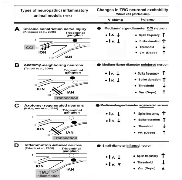
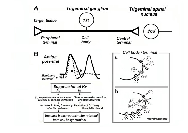
Previous studies in several different trigeminal nerve injury/inflammation models indicated that the hyperexcitability of primary afferent neurons contributes to the pain pathway underlying mechanical allodynia. Although multiple types of voltage-gated ion channels are associated with neuronal hyperexcitability, voltage-gated K+ channels (Kv) are one of the important physiological regulators of membrane potentials in excitable tissues, including nociceptive sensory neurons. Since the opening of K+ channels leads to hyperpolarization of cell membrane and a consequent decrease in cell excitability, several Kv channels have been proposed as potential target candidates for pain therapy. In this review, we focus on common changes measured in the Kv channels of several different trigeminal neuropathic/inflammatory pain animal models, particularly the relationship between changes in Kv channels and the excitability of trigeminal ganglion (TRG) neurons. We also discuss the potential of Kv channel openers as therapeutic agents for trigeminal neuropathic/inflammatory pain, such as mechanical allodynia.


Figures

Figure 1
Figure 1
Trigeminal neuropathic/inflammatory pain models and changes in TRG neuronal activities. A: Chronic constriction nerve injury (CCI) model. Three days after infraorbital nerve (ION) chronic constriction. Target trigeminal ganglion (TRG) neurons were labeled by fluorogold (FG). Whole-cell patch-clamp recordings under current-/voltage-clamp configurations were performed on medium/large-diameter CCI neurons. B: Axotomy-neighboring neuron model. In this model, we found mechanical allodynia in the territory of ION at 2 days after inferior alveolar nerve (IAN) transection. Uninjured TRG neurons innervating the territory of ION were labeled by FG and patch-clamp recordings under current-/voltage-clamp configuration was performed on medium-/large-diameter neurons. C: Axotomy-regenerated neuron model. In this model, we observed mechanical allodynia at 14 days after IAN transection. FG-injection at 14 days after IAN transection showed massive labeling of trigeminal ganglion containing FG-labeled small/large-diameter neurons and the patch-clamp recordings under current-/voltage-clamp configuration indicated axotomy-regenerated neurons. D: Inflammation model. Inflammation was induced by injection of CFA into the rat temporomandibular joint (TMJ). We found mechanical allodynia in the territory of ION at 2 days after CFA injection. In FG-labeled small-diameter TRG neurons innervating TMJ, the whole-cell patch-clamp recordings under current-/voltage-clamp configurations were performed on inflammatory neurons. Blackened circles indicate the trigeminal ganglion neurons. I-III: trigeminal nerves, IA: transient K current, IK: sustained K currents, ↑: increase, ↓: decrease, ▼: tendency for decrease in IK, but not significant, ▲: tendency for depolarization of membrane potential (Vm), but not significant, Depo: Depolarization, V-clamp : Voltage-clamp, I-clamp: Current-clamp.
Figure 2
Figure 2
Relationship between depression of Kv and excitability of TRG neurons under trigeminal neuropathic/inflammatory conditions. A: The operational component of primary sensory neurons (1st) includes a peripheral terminal that innervates target tissues and transduces sensory stimuli. An axon conducts action potentials from the periphery to the central nervous system via the cell body in the trigeminal ganglion and central terminal, from where information is transferred to second order neurons (2nd) at the central synapse (e.g., trigeminal spinal nucleus). B: The relationship between the depression of Kv channels and the excitability of TRG neurons. The increases in action potential firing and duration prolongs the opening of voltage-gated Ca2+ channels (Cav), potentiating Ca2+ influx, and causing an increase in neurotransmitter released from the cell bodies and nerve terminals. These changes can alter the properties of trigeminal pain pathways.

References

    1. Devor M. Strategies for finding new pharmacological targets for neuropathic pain. Clin Pain Head Reports. 2004;8:187–191. doi: 10.1007/s11916-004-0050-8. - DOI - PubMed
    1. Niv D, Devor M. Refractory neuropathic pain: the nature and extent of the problem. Pain Prac. 2006;6:3–9. doi: 10.1111/j.1533-2500.2006.00052.x. - DOI - PubMed
    1. Schwartz J, Naff N. The management of neuropathic pain. Neurosurg Clin N Am. 2004;15:231–239. doi: 10.1016/j.nec.2004.01.001. - DOI - PubMed
    1. Scholtz J, Woolf CJ. Can we conquer pain? Nat Neurosci. 2002;5:1062–1067. doi: 10.1038/nn942. - DOI - PubMed
    1. Costigas M, Scholz J, Woolf CJ. Neuropathic pain: A maladaptive response of nervous system to damage. Annu Rev Neurosci. 2009;32:1–32. doi: 10.1146/annurev.neuro.051508.135531. - DOI - PMC - PubMed

Substances

LinkOut - more resources