. 2011 Jan 13;69(1):33-43.
doi: 10.1016/j.neuron.2010.12.002.

Dysregulation of limbic and auditory networks in tinnitus

Affiliations

Dysregulation of limbic and auditory networks in tinnitus

Amber M Leaver et al. Neuron. .

Abstract

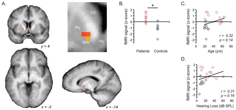
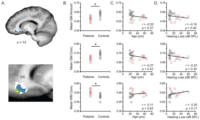
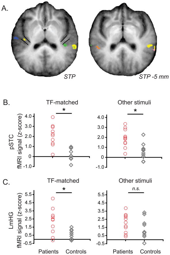
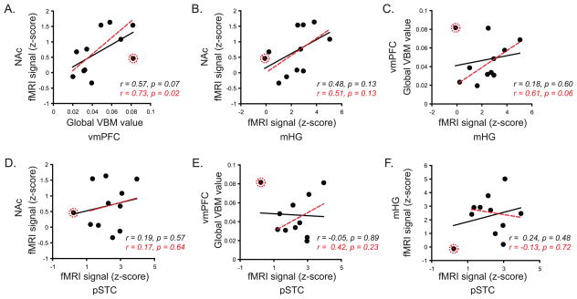
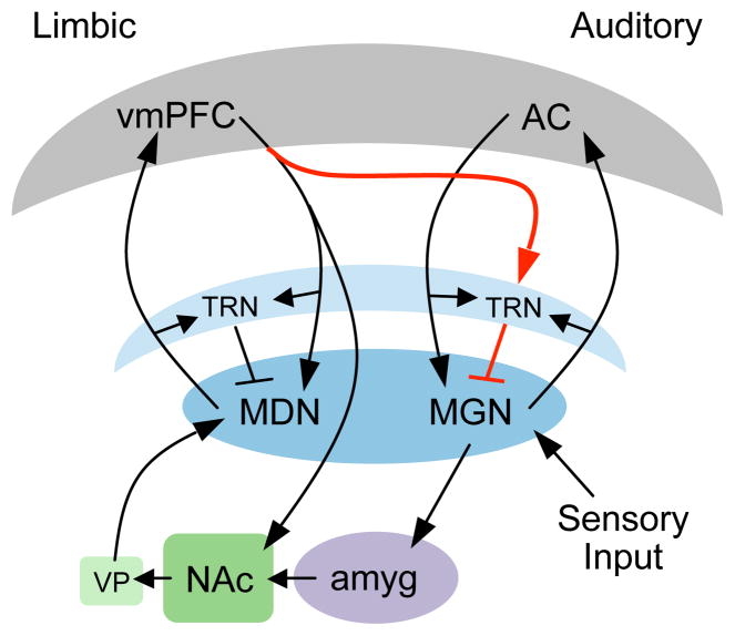
Tinnitus is a common disorder characterized by ringing in the ear in the absence of sound. Converging evidence suggests that tinnitus pathophysiology involves damage to peripheral and/or central auditory pathways. However, whether auditory system dysfunction is sufficient to explain chronic tinnitus is unclear, especially in light of evidence implicating other networks, including the limbic system. Using functional magnetic resonance imaging and voxel-based morphometry, we assessed tinnitus-related functional and anatomical anomalies in auditory and limbic networks. Moderate hyperactivity was present in the primary and posterior auditory cortices of tinnitus patients. However, the nucleus accumbens exhibited the greatest degree of hyperactivity, specifically to sounds frequency-matched to patients' tinnitus. Complementary structural differences were identified in ventromedial prefrontal cortex, another limbic structure heavily connected to the nucleus accumbens. Furthermore, tinnitus-related anomalies were intercorrelated in the two limbic regions and between limbic and primary auditory areas, indicating the importance of auditory-limbic interactions in tinnitus.


Figures

Figure 1
Figure 1
Hyperactivity in tinnitus patients was localized to the ventral striatum near the nucleus accumbens (center of gravity: X,Y,Z = −16, 6, −0.5; volume = 108 mm3). A. Voxels exhibiting significant (p(corr) < 0.05) between-groups differences in fMRI signal are shown on group-averaged anatomical images. Inset in A shows a close-up of the coronal image, emphasizing the position of the cluster in the ventral striatum (nucleus accumbens, NAc; caudate, Cd; putamen, Pu; hypothalamus, Hy). B–D. Mean fMRI signal for each subject is plotted for tinnitus patients (red circles) and stimulus-matched control participants (grey diamonds) in B. A black circle marks the tinnitus patient reporting comorbid depression; color scheme is constant throughout. Asterisk denotes statistical significance at the single-voxel level demonstrated in A. This functional difference in NAc is not related to participant age (C) or mean hearing loss (D). Note that where tinnitus patients overlap with control participants in age and mean hearing loss, NAc response still exhibits a clear between-groups difference.
Figure 2
Figure 2
In a masked analysis restricted to auditory cortex and thalamus, hyperactivity in tinnitus patients was demonstrated in auditory cortex. A. Voxels that demonstrated between-groups differences in fMRI signal (p < 0.01, k > 108 mm3) are shown on group-averaged anatomical images, rotated to visualize the superior temporal plane (STP). Tinnitus-related hyperactivity was seen during trials containing TF-matched stimuli (yellow), stimuli less than 0.5 octaves below the TF (green), less than 0.5 octaves above the TF (orange), and more than 0.5 octaves above the TF (pink). Blue marks a single instance where signal was less for tinnitus patients. B–C. Mean fMRI signal is plotted for tinnitus patients (red) and control participants (grey) for TF-matched stimuli and other stimuli in pSTC (B) and mHG (C). Brain activity in patients during TF-matched trials was also significantly greater than control participants’ during non-TF-matched trials in these regions (pSTC: t(20) = 4.09, p = 0.0003; mHG: t(20) = 1.68, p = 0.05; one-tailed tests).
Figure 3
Figure 3
Structural differences between tinnitus patients and control participants were identified in ventromedial prefrontal cortex (vmPFC). A. Voxels demonstrating significant differences in VBM values between groups are shown on group-averaged anatomical images. Inset in A is a close-up of the sagittal image, showing the position of anatomical differences located in vmPFC inferior to the corpus callosum (CC). The position of basal ganglia structures is also indicated (caudate, CD; nucleus accumbens, NAc). Between-groups differences were seen in modulated and unmodulated grey matter (GMm and GMum, respectively) and modulated white matter (WMm) images. White corresponds to WMm differences, yellow marks GMm and WMm differences, blue marks GMum and WMm differences, and green marks differences in GMm, GMum, and WMm. B–C. Mean VBM values are plotted for each tinnitus patient (red circles) and control (grey diamonds). Asterisks in B denote the statistically significant differences in GMm (amount, top), GMum (concentration, middle), and WMm (amount, bottom) at the single-voxel level shown in A. These differences were not related to age (C) or mean hearing loss (D).
Figure 4
Figure 4
Correlations between functional and anatomical markers are displayed. Data corresponding to NAc, mHG, and pSTC reflect fMRI signal during TF-matched trials. Global VBM values in vmPFC reflect the mean difference in modulated and unmodulated grey matter and modulated white matter from the corresponding mean values in control participants. Thus, large global VBM values indicate larger difference from controls, while smaller values indicate smaller tinnitus-related differences. A single outlier (see Methods) is marked in red; r and p values are displayed for each pairwise correlation both including (black) and excluding (red) this outlier.
Figure 5
Figure 5
Schematic of proposed auditory-limbic interactions in tinnitus. Sensory input originates subcortically and enters both auditory and limbic circuits via the medial geniculate nucleus (MGN). Under normal circumstances, the limbic system may identify a sensory signal as perceptually irrelevant (e.g., transient tinnitus following loud noise exposure), and inhibit the unwanted signal at the MGN via projections from the ventromedial prefrontal cortex (vmPFC) to the auditory thalamic reticular nucleus (TRN, red pathway). Thus, propagation of the unwanted signal (e.g., transient tinnitus) is reduced in both circuits. In chronic tinnitus, inefficient vmPFC output prevents inhibition of the tinnitus signal, resulting in continued thalamocortical activity and the constant perceptual presence of the tinnitus signal. Cortical structures are noted in grey, thalamus is noted in blue, basal ganglia in green, and amygdala in lavender. Schematic is not to scale, and position of structures was not made to accurately reflect anatomical position. Abbreviations: medial dorsal nucleus (MDN), ventral pallidum (VP), amygdala (amyg), auditory cortex (AC).

References

    1. Ahmed SH, Koob GF. Transition from moderate to excessive drug intake: change in hedonic set point. Science. 1998;282:298–300. - PubMed
    1. Alain C, Reinke K, McDonald KL, Chau W, Tam F, Pacurar A, Graham S. Left thalamo-cortical network implicated in successful speech separation and identification. Neuroimage. 2005;26:592–599. - PubMed
    1. Ashburner J, Friston KJ. Voxel-based morphometry--the methods. Neuroimage. 2000;11:805–821. - PubMed
    1. Bar M. A cognitive neuroscience hypothesis of mood and depression. Trends Cogn Sci. 2009;13:456–463. - PMC - PubMed
    1. Blood AJ, Zatorre RJ, Bermudez P, Evans AC. Emotional responses to pleasant and unpleasant music correlate with activity in paralimbic brain regions. Nat Neurosci. 1999;2:382–387. - PubMed

Publication types