. 2011 Feb 22;108(8):3193-8.
doi: 10.1073/pnas.1012464108. Epub 2011 Jan 10.

Masking the 5' terminal nucleotides of the hepatitis C virus genome by an unconventional microRNA-target RNA complex

Affiliations

Masking the 5' terminal nucleotides of the hepatitis C virus genome by an unconventional microRNA-target RNA complex

Erica S Machlin et al. Proc Natl Acad Sci U S A. .

Abstract

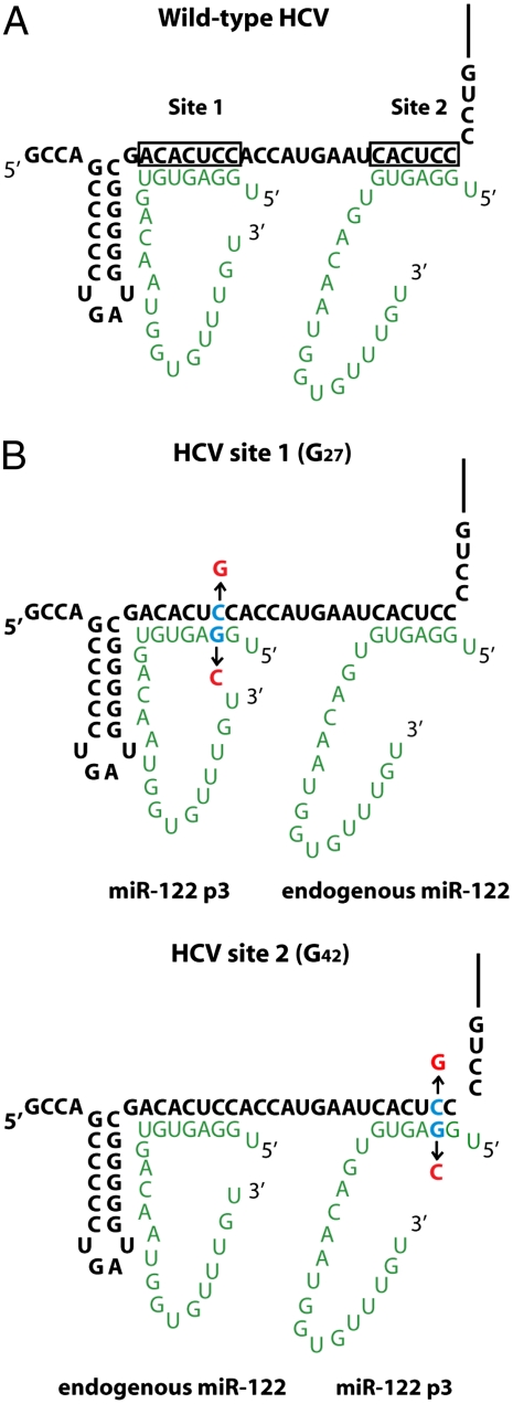
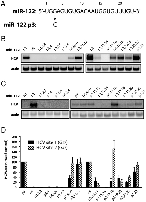
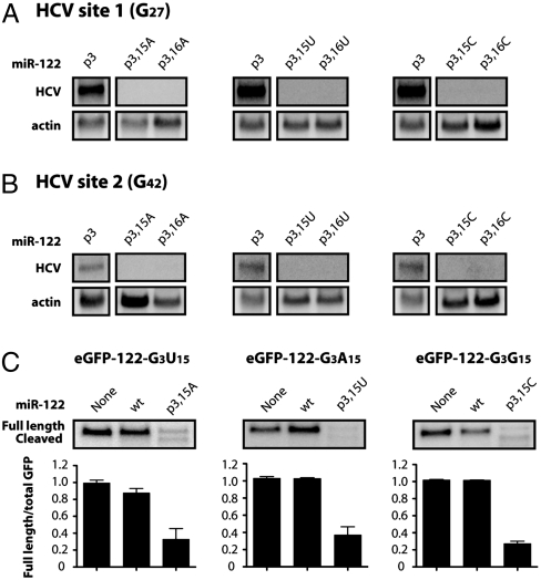
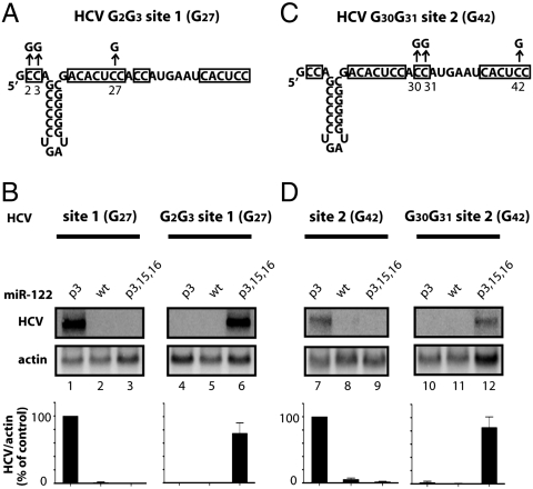
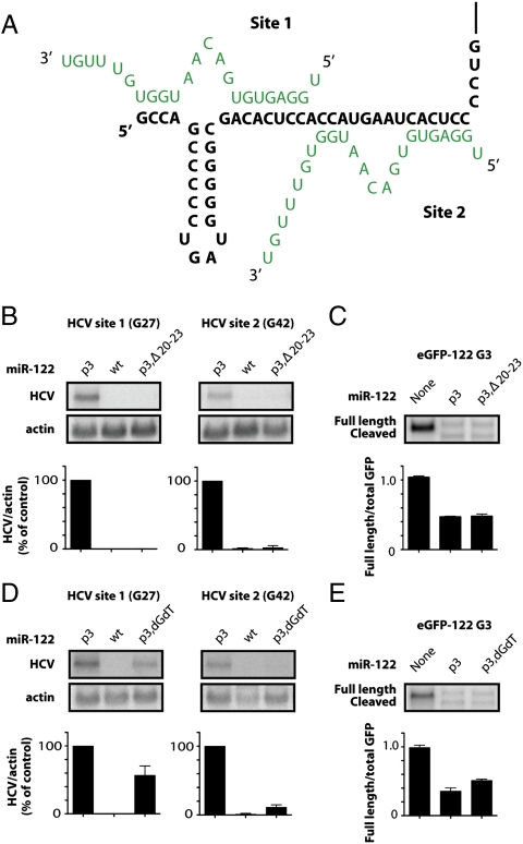
Hepatitis C virus subverts liver-specific microRNA, miR-122, to upregulate viral RNA abundance in both infected cultured cells and in the liver of infected chimpanzees. These findings have identified miR-122 as an attractive antiviral target. Thus, it is imperative to know whether a distinct functional complex exists between miR-122 and the viral RNA versus its normal cellular target mRNAs. Toward this goal, effects on viral RNA abundance of mutated miR-122 duplex molecules, bound at each of the two target sites in the viral genome, were compared to effects on microRNA- or siRNA-mediated regulation of reporter target mRNAs. It was found that miR-122 formed an unusual microRNA complex with the viral RNA that is distinct from miR-122 complexes with reporter mRNAs. Notably, miR-122 forms an oligomeric complex in which one miR-122 molecule binds to the 5' terminus of the hepatitis C virus (HCV) RNA with 3' overhanging nucleotides, masking the 5' terminal sequences of the HCV genome. Furthermore, specific internal nucleotides as well as the 3' terminal nucleotides in miR-122 were absolutely required for maintaining HCV RNA abundance but not for microRNA function. Both miR-122 molecules utilize similar internal nucleotides to interact with the viral genome, creating a bulge and tail in the miR-122 molecules, revealing tandemly oriented oligomeric RNA complexes. These findings suggest that miR-122 protects the 5' terminal viral sequences from nucleolytic degradation or from inducing innate immune responses to the RNA terminus. Finally, this remarkable microRNA-mRNA complex could be targeted with compounds that inactivate miR-122 or interfere with this unique RNA structure.


Conflict of interest statement

The authors declare no conflict of interest.

Figures

Fig. 1.
Fig. 1.
Interactions between miR-122 (green) and HCV (black). (A) Interactions of two miR-122 molecules with HCV RNA. Nucleotides (nts) 2–8 of miR-122 interact at site 1 and nts 2–7 of miR-122 interact at site 2 with the 5′ end of HCV RNA. (B) Location of introduced nucleotide substitutions for stepwise mutational analyses. Mutant miR-122 molecules are directed to HCV site 1 via HCV site 1 (G27), or to HCV site 2 via HCV site 2 (G42). Mutated nucleotides in HCV RNA or miR-122 are shown in red. Endogenous miR-122 binds only to wild-type seed sites in HCV.
Fig. 2.
Fig. 2.
Effects of HCV site 1- or HCV site 2-bound miR-122 molecules on HCV RNA abundance. (A) Sequence of wild-type miR-122 and miR-122 p3. Position 3 of miR-122 was mutated from a guanosine to a cytidine to produce miR-122 p3. (B) HCV site 1 (G27) RNA accumulation was measured by Northern blot analysis 5 d postelectroporation in Huh-7 cells. MiR-122 p3 duplexes, mutated in two nucleotide increments, were transfected into cells 1 d prior to, and 1 and 3 d postelectroporation. Nucleotides were mutated to their Watson–Crick base pair. (C) HCV site 2 (G42) RNA and miR-122 mutants were expressed and analyzed as in B. (D) Quantitation of HCV RNA levels. HCV RNA abundances were normalized to γ-actin levels. Data from cells transfected with miR-122 p3 were set to 100%. The data shown represent at least three independent replicates. Error bars represent standard error of the mean.
Fig. 3.
Fig. 3.
Effects of nucleotide 15 or 16 in miR-122 on HCV RNA abundance (A) and siRNA-mediated cleavage of GFP reporter RNA (B). (A) Position 15 or 16 in miR-122 p3 was mutated to A, U, or C, and the mutants were tested in HCV site 1 (G27) RNA electroporation assays as described in Fig. 2. Northern blots representative of at least three independent replicates are shown. (B) HCV site 2 (G42) RNA and miR-122 mutants were expressed and analyzed as in A. (C) MiR-122 p3 duplexes containing an A, U, or C at position 15 were cotransfected into HeLa cells with a plasmid expressing GFP that contained a perfectly complementary site to the mutant miR-122 molecule in its 3′ noncoding region. GFP mRNA was measured by Northern blot analysis. Full length and cleaved GFP mRNA are indicated. Quantitation is shown as the ratio of full length GFP mRNA to total GFP mRNA and represents at least three independent replicates. Error bars represent standard error of the mean.
Fig. 4.
Fig. 4.
Interaction of miR-122 nucleotides 15 and 16 with HCV RNA. (A) Diagram of HCV G2G3 site 1 (G27) RNA that was mutated to include additional C to G mutations at nucleotides 2 and 3. (B) HCV site 1 (G27) and HCV G2G3 site 1 G27) RNA abundance in cells transfected with miR-122 p3, WT, or p3,15,16 duplexes, as described in Fig. 2. Northern blot data are representative of at least three independent replicates. Error bars represent standard error of the mean. (C) Diagram of HCV G30G31 site 2 (G42) RNA that was mutated to include additional C to G mutations at nucleotides 30 and 31. (D) HCV site 2 (G42) RNA, HCV G30G31 site 2 (G42), and miR-122 mutants were expressed and analyzed as in Fig. 2.
Fig. 5.
Fig. 5.
Effects of 3′ end sequences of miR-122 on HCV RNA abundance. (A) Model for miR-122 (green)-HCV (black) interactions at the 5′ end of the HCV genome. (B) Effects of 3′ end deletions in miR-122. A 19-nucleotide-long miR-122 p3 duplex (p3,Δ20–23) was targeted to site 1 or 2 in HCV RNA electroporation assays as described in Fig. 2. (C) siRNA-mediated GFP cleavage assays in HeLa cells transfected with p3,Δ20–23 as described in Fig. 3. (D) Effects of 3′ end nucleotide compositions in miR-122. The last two nucleotides of miR-122 p3 were replaced with deoxynucleotides dG and dT (p3,dGdT) and targeted to site 1 or 2 in HCV RNA electroporation assays as described in Fig. 2. (E) siRNA-mediated GFP cleavage assays in HeLa cells transfected with p3,dGdT as described in Fig. 3.

Comment in

  • End game: getting the most out of microRNAs.
    You S, Murray CL, Luna JM, Rice CM. You S, et al. Proc Natl Acad Sci U S A. 2011 Feb 22;108(8):3101-2. doi: 10.1073/pnas.1019613108. Epub 2011 Feb 9. Proc Natl Acad Sci U S A. 2011. PMID: 21307312 Free PMC article. No abstract available.

References

    1. Moradpour D, Penin F, Rice CM. Replication of hepatitis C virus. Nat Rev Microbiol. 2007;5:453–463. - PubMed
    1. Hoofnagle JH. Course and outcome of hepatitis C. Hepatology. 2002;36:S21–S29. - PubMed
    1. Tellinghuisen TL, Evans MJ, von Hahn T, You S, Rice CM. Studying hepatitis C virus: Making the best of a bad virus. J Virol. 2007;81:8853–8867. - PMC - PubMed
    1. Jopling CL, Yi M, Lancaster AM, Lemon SM, Sarnow P. Modulation of hepatitis C virus RNA abundance by a liver-specific MicroRNA. Science. 2005;309:1577–1581. - PubMed
    1. Jopling CL, Schutz S, Sarnow P. Position-dependent function for a tandem microRNA miR-122-binding site located in the hepatitis C virus RNA genome. Cell Host Microbe. 2008;4:77–85. - PMC - PubMed

Publication types