

Thyrotrophin receptor signaling dependence of Braf-induced thyroid tumor initiation in mice
- PMID: 21220306
- PMCID: PMC3029699
- DOI: 10.1073/pnas.1015557108
Thyrotrophin receptor signaling dependence of Braf-induced thyroid tumor initiation in mice
Abstract
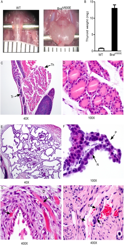
Mutations of BRAF are found in ∼45% of papillary thyroid cancers and are enriched in tumors with more aggressive properties. We developed mice with a thyroid-specific knock-in of oncogenic Braf (LSL-Braf(V600E)/TPO-Cre) to explore the role of endogenous expression of this oncoprotein on tumor initiation and progression. In contrast to other Braf-induced mouse models of tumorigenesis (i.e., melanomas and lung), in which knock-in of Braf(V600E) induces mostly benign lesions, Braf-expressing thyrocytes become transformed and progress to invasive carcinomas with a very short latency, a process that is dampened by treatment with an allosteric MEK inhibitor. These mice also become profoundly hypothyroid due to deregulation of genes involved in thyroid hormone biosynthesis and consequently have high TSH levels. To determine whether TSH signaling cooperates with oncogenic Braf in this process, we first crossed LSL-Braf(V600E)/TPO-Cre with TshR knockout mice. Although oncogenic Braf was appropriately activated in thyroid follicular cells of these mice, they had a lower mitotic index and were not transformed. Thyroid-specific deletion of the Gsα gene in LSL-Braf(V600E)/TPO-Cre/Gnas-E1(fl/fl) mice also resulted in an attenuated cancer phenotype, indicating that the cooperation of TshR with oncogenic Braf is mediated in part by cAMP signaling. Once tumors were established in mice with wild-type TshR, suppression of TSH did not revert the phenotype. These data demonstrate the key role of TSH signaling in Braf-induced papillary thyroid cancer initiation and provide experimental support for recent observations in humans pointing to a strong association between TSH levels and thyroid cancer incidence.
Conflict of interest statement
The authors declare no conflict of interest.
Figures


Similar articles
-
Thyrotropin signaling confers more aggressive features with higher genomic instability on BRAF(V600E)-induced thyroid tumors in a mouse model.Thyroid. 2014 Mar;24(3):502-10. doi: 10.1089/thy.2013.0038. Epub 2014 Jan 15. Thyroid. 2014. PMID: 23924149 Free PMC article.
-
TSH overcomes Braf(V600E)-induced senescence to promote tumor progression via downregulation of p53 expression in papillary thyroid cancer.Oncogene. 2016 Apr 14;35(15):1909-18. doi: 10.1038/onc.2015.253. Epub 2015 Oct 19. Oncogene. 2016. PMID: 26477313 Free PMC article.
-
Postnatal expression of BRAFV600E does not induce thyroid cancer in mouse models of thyroid papillary carcinoma.Endocrinology. 2013 Nov;154(11):4423-30. doi: 10.1210/en.2013-1174. Epub 2013 Aug 22. Endocrinology. 2013. PMID: 23970782
-
The role of BRAF in the pathogenesis of thyroid carcinoma.Front Biosci (Landmark Ed). 2015 Jun 1;20(7):1068-78. doi: 10.2741/4359. Front Biosci (Landmark Ed). 2015. PMID: 25961545 Review.
-
Targeting BRAF in thyroid cancer.Br J Cancer. 2007 Jan 15;96(1):16-20. doi: 10.1038/sj.bjc.6603520. Epub 2006 Dec 19. Br J Cancer. 2007. PMID: 17179987 Free PMC article. Review.
Cited by
-
Cancer Stem Cells in Thyroid Tumors: From the Origin to Metastasis.Front Endocrinol (Lausanne). 2020 Aug 25;11:566. doi: 10.3389/fendo.2020.00566. eCollection 2020. Front Endocrinol (Lausanne). 2020. PMID: 32982967 Free PMC article. Review.
-
Relationship Between Thyroid-Stimulating Hormone Level and Aggressive Pathological Features of Papillary Thyroid Cancer.Sisli Etfal Hastan Tip Bul. 2022 Mar 28;56(1):126-131. doi: 10.14744/SEMB.2022.14554. eCollection 2022. Sisli Etfal Hastan Tip Bul. 2022. PMID: 35515965 Free PMC article.
-
Mouse Model of Poorly Differentiated Thyroid Carcinoma Driven by STRN-ALK Fusion.Am J Pathol. 2018 Nov;188(11):2653-2661. doi: 10.1016/j.ajpath.2018.07.012. Epub 2018 Aug 18. Am J Pathol. 2018. PMID: 30125543 Free PMC article.
-
Clinicopathological Profile of Thyroid Carcinoma in Young Patients: An Indonesian Single-Center Study.J Thyroid Res. 2022 Jan 11;2022:9944083. doi: 10.1155/2022/9944083. eCollection 2022. J Thyroid Res. 2022. PMID: 35059180 Free PMC article.
-
Oncogenic BRAF disrupts thyroid morphogenesis and function via twist expression.Elife. 2017 Mar 28;6:e20728. doi: 10.7554/eLife.20728. Elife. 2017. PMID: 28350298 Free PMC article.
References
-
- Tramontano D, Cushing GW, Moses AC, Ingbar SH. Insulin-like growth factor-I stimulates the growth of rat thyroid cells in culture and synergizes the stimulation of DNA synthesis induced by TSH and Graves’-IgG. Endocrinology. 1986;119:940–942. - PubMed
-
- Kimura T, et al. Regulation of thyroid cell proliferation by TSH and other factors: A critical evaluation of in vitro models. Endocr Rev. 2001;22:631–656. - PubMed
-
- Roger PP, Servais P, Dumont JE. Stimulation by thyrotropin and cyclic AMP of the proliferation of quiescent canine thyroid cells cultured in a defined medium containing insulin. FEBS Lett. 1983;157:323–329. - PubMed
-
- Parma J, et al. Somatic mutations in the thyrotropin receptor gene cause hyperfunctioning thyroid adenomas. Nature. 1993;365:649–651. - PubMed
-
- Lyons J, et al. Two G protein oncogenes in human endocrine tumors. Science. 1990;249:655–659. - PubMed
Publication types
MeSH terms
Substances
Grants and funding
- R01 CA072597/CA/NCI NIH HHS/United States
- R37 DK015070/DK/NIDDK NIH HHS/United States
- P30 CA08748/CA/NCI NIH HHS/United States
- P30 CA008748/CA/NCI NIH HHS/United States
- ImNIH/Intramural NIH HHS/United States
- 13083/CRUK_/Cancer Research UK/United Kingdom
- R24 CA83084/CA/NCI NIH HHS/United States
- R24 CA083084/CA/NCI NIH HHS/United States
- F32CA136178/CA/NCI NIH HHS/United States
- DK17050/DK/NIDDK NIH HHS/United States
- P50-CA92629/CA/NCI NIH HHS/United States
- R01 CA050706/CA/NCI NIH HHS/United States
- CA72597/CA/NCI NIH HHS/United States
- P50 CA092629/CA/NCI NIH HHS/United States
- CA50706/CA/NCI NIH HHS/United States
- F32 CA136178/CA/NCI NIH HHS/United States
LinkOut - more resources
Full Text Sources
Other Literature Sources
Medical
Molecular Biology Databases
Research Materials
