. 2011 Jan 25;108(4):1415-20.
doi: 10.1073/pnas.1011275108. Epub 2011 Jan 10.

Phosphorylation of glutamyl-prolyl tRNA synthetase by cyclin-dependent kinase 5 dictates transcript-selective translational control

Affiliations

Phosphorylation of glutamyl-prolyl tRNA synthetase by cyclin-dependent kinase 5 dictates transcript-selective translational control

Abul Arif et al. Proc Natl Acad Sci U S A. .

Abstract

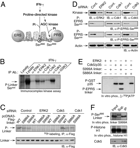
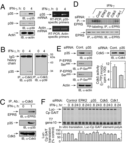
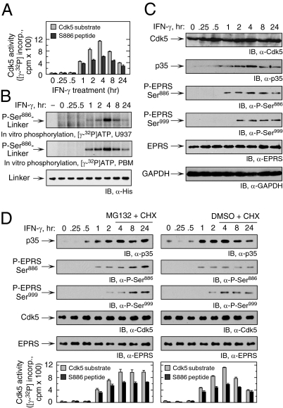
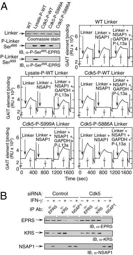
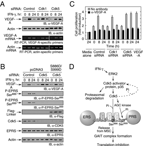
Cyclin-dependent kinase 5 (Cdk5) is an atypical but essential member of the Cdk kinase family, and its dysregulation or deletion has been implicated in inflammation-related disorders by an undefined mechanism. Here we show that Cdk5 is an indispensable activator of the GAIT (IFN-γ-activated inhibitor of translation) pathway, which suppresses expression of a posttranscriptional regulon of proinflammatory genes in myeloid cells. Through induction of its regulatory protein, Cdk5R1 (p35), IFN-γ activates Cdk5 to phosphorylate Ser(886) in the linker domain of glutamyl-prolyl tRNA synthetase (EPRS), the initial event in assembly of the GAIT complex. Cdk5/p35 also induces, albeit indirectly via a distinct kinase, phosphorylation of Ser(999), the second essential event in GAIT pathway activation. Diphosphorylated EPRS is released from its residence in the tRNA multisynthetase complex for immediate binding to NS1-associated protein and subsequent binding to ribosomal protein L13a and GAPDH. The mature heterotetrameric GAIT complex binds the 3' UTR GAIT element of VEGF-A and other target mRNAs and suppresses their translation in myeloid cells. Inhibition of Cdk5/p35 inhibits both EPRS phosphorylation events, prevents EPRS release from the tRNA multisynthetase complex, and blocks translational suppression of GAIT element-bearing mRNAs, resulting in increased expression of inflammatory proteins. Our study reveals a unique role of Cdk5/p35 in activation of the major noncanonical function of EPRS, namely translational control of macrophage inflammatory gene expression.


Conflict of interest statement

The authors declare no conflict of interest.

Figures

Fig. 1.
Fig. 1.
ERK2 and Cdk5 are required for phosphorylation of EPRS at Ser886. (A) Schematic of the IFN-γ–activated kinase pathway directing the phosphorylation of human EPRS at Ser886 and Ser999. (B) Immunocomplexes containing ERK2, Cdk1, and Cdk5 phosphorylate EPRS at Ser886. U937 cells were treated for 4 h with IFN-γ, and candidate proline-directed kinases were immunoprecipitated from lysates. The immunocomplexes were used to phosphorylate recombinant EPRS linker bearing a Ser999-to-Ala mutation in the presence of [γ-32P]ATP. (C) ERK2- and Cdk5-knockdown block 32P incorporation into EPRS linker. U937 cells were cotransfected with Flag-tagged EPRS linker (WT), Ser886-to-Ala (S886A) and Ser999-to-Ala (S999A) mutants, and siRNAs (or control) against ERK2, Cdk5, and Cdk1. Transfected cells were incubated for 4 h with 32P-orthophosphate and IFN-γ. Lysates were immunoprecipitated with anti-Flag antibody, and EPRS linker phosphorylation was determined by autoradiography (Upper). Total transfected EPRS linker was determined by immunoblot analysis (IB) with anti-Flag antibody (Lower). (D) ERK2 and Cdk5 are required for phosphorylation of endogenous EPRS. U937 cells were transfected with the siRNAs shown. Scrambled siRNAs were used as controls (Cont). After 24 h of recovery, cells were treated with IFN-γ for an additional 4 h. Phosphospecific antibodies were used to detect Ser886 and Ser999 phosphorylation. Immunoblot analysis with anti-actin antibody served as a loading control. (E) Recombinant Cdk5 phosphorylates EPRS at Ser886. Recombinant ERK2 and CDK5 activated by GST-tagged p35 were used for in vitro phosphorylation of recombinant EPRS linker substrates bearing either a S999A (Ser886 substrate) or S886A (Ser999 substrate) mutation. (F) Phosphorylation of EPRS linker at Ser886 by endogenous Cdk5. Cdk5 from IFN-γ–activated U937 cells was immunoprecipitated with anti-Cdk5 antibody. Lysate (input), immunoprecipitated fraction (IP), and supernatant (Sup) were used for in vitro phosphorylation of recombinant S999A linker (Top) and histone H1 (Middle) substrates. Cdk5 was detected with anti-Cdk5 antibody (Bottom).
Fig. 2.
Fig. 2.
Induction of p35 activator protein by IFN-γ is required for Ser886 phosphorylation by Cdk5. (A) IFN-γ induces p35 activator protein. (Left) Lysates from U937 cells treated with IFN-γ for 4 h were immunoblotted. (Right) p35 and β-actin mRNA was determined by RT-PCR of total cellular RNA. (B) IFN-γ induces Cdk5 binding to p35. Lysates from U937 cells treated with IFN-γ for 4 h were coimmunoprecipitated with anti-Cdk5 antibody and probed with anti-p35 (Left) and anti-p39 (Right) antibodies. (C) Cdk5/p35 phosphorylates EPRS at Ser886. Lysates from U937 cells treated with IFN-γ for 1 h were immunoprecipitated and immunoblotted as shown. (D) Knockdown of p35 blocks EPRS phosphorylation. (Upper) siRNAs were transfected into U937 cells and incubated with 32P-orthophosphate and IFN-γ for 4 h. Lysates were immunoprecipitated with anti-EPRS antibody, and EPRS phosphorylation was determined by autoradiography. (Lower) Total EPRS was detected with anti-EPRS antibody. (E) Knockdown of p35 activator protein blocks two-site EPRS phosphorylation. U937 cells were transfected with siRNA against p35 and then treated with IFN-γ as in Fig. 1D. (Left) Knockdown efficiency and EPRS phosphorylation were determined by immunoblot analysis.(Right) Lysates were incubated with anti-Cdk5 antibody, and precipitates were used for in vitro phosphorylation of Cdk5-specific peptide substrate. Aliquots were spotted on phosphocellulose P-81, and the incorporation of 32P into peptide was determined by scintillation counting (mean ± SEM; n = 3 experiments). (F) ERK2, p35, and Cdk5 are required for GAIT complex–mediated translational suppression. U937 cells were transfected with the siRNAs shown and incubated with IFN-γ for up to 24 h. Lysates were added to RRL for in vitro translation of capped, polyadenylated luciferase (Luc) reporter transcript bearing the Cp GAIT element in the presence of 35S-Met. T7 gene 10 RNA lacking the GAIT element was cotranslated as a specificity control. Luc translation was quantified by densitometry, normalized by T7 gene 10, and expressed as percentage of control (mean ± SEM; n = 3 experiments).
Fig. 3.
Fig. 3.
Temporal correspondence of IFN-γ–stimulated p35 expression, Cdk5 activation, and EPRS phosphorylation. (A) IFN-γ–mediated induction of Cdk5 activity is maximal at 4 h. Lysates from U937 cells treated with IFN-γ for up to 24 h were incubated with anti-Cdk5 antibody, and the precipitates were used for in vitro phosphorylation of Cdk5-specific (gray bars) or Ser886-containing EPRS (black bars) peptide substrates (mean ± SEM; n = 3 experiments). (B) Lysates of IFN-γ–treated U937 cells (Top) or PBMs (Middle) were used for in vitro phosphorylation of recombinant His-tagged EPRS linker containing a Ser999-to-Ala mutation that permits only Ser886 phosphorylation. Immunoblot analysis with anti-His antibody served as a loading control (Bottom). (C) IFN-γ induces p35 expression. Lysates from IFN-γ–treated U937 cells were immunoblotted as shown. (D) IFN-γ–induced p35 expression is down-regulated by proteasomal degradation. U937 cells were pretreated with IFN-γ for 4 h and then with IFN-γ, MG132 (10 μM), and cycloheximide (CHX; 30 μg/mL) for up to 24 h (Left). Control cells were treated with DMSO and CHX (Right). Lysate expression of p35, Cdk5, and phosphorylated and total EPRS was determined by immunoblot analysis. Cdk5 activity was determined in lysates after precipitation with anti-Cdk5 antibody as described in Fig. 2E.
Fig. 4.
Fig. 4.
Cdk5-mediated EPRS phosphorylation is required for the release of EPRS from the MSC and for binding to GAIT element RNA. (A) EPRS linker phosphorylated at Ser886 by Cdk5 reconstitutes the GAIT complex assembly and binding to GAIT element RNA. His-tagged, WT, and mutant (S886A and S999A) EPRS linker proteins were phosphorylated by incubation with lysate from IFN-γ–treated U937 cells or with recombinant Cdk5/p35, and then repurified using Ni-affinity resin (Top, Left). Specific phosphorylation was confirmed using anti-phosphospecific antibodies. Unmodified (Top, Right), lysate-phosphorylated (Middle, Left), or Cdk5-phosphorylated WT (Middle, Right), S999A (Bottom, Left), and S886A (Bottom, Right) EPRS linkers were preincubated with NSAP1 or with NSAP1, GAPDH, and phospho-L13a (P-L13a), and binding to biotinylated Cp GAIT element RNA immobilized on a streptavidin sensor chip was determined by SPR and expressed as resonance units (RU). (B) Cdk5-mediated phosphorylation induces EPRS release from MSC. U937 cells were transfected with siRNA targeting Cdk5 (and control siRNA) and then treated with IFN-γ for 4 h. Lysates were immunoprecipitated with antibodies against EPRS, LysRS (KRS, MSC constituent), and NSAP1 (pre-GAIT complex constituent), and evaluated by immunoblot analysis.
Fig. 5.
Fig. 5.
Cdk5-mediated EPRS phosphorylation regulates VEGF-A expression. (A) Inhibition of Cdk5 expression enhances VEGF-A expression. U937 cells were transfected with siRNA as shown and then incubated with IFN-γ for up to 24 h. (Upper) Lysates were probed with anti–VEGF-A or actin antibodies. (Lower) RT-PCR analysis of total cellular RNA was performed using gene-specific primers. (B) Expression of phosphomimetic EPRS linker in Cdk5 knockdown cells restores inhibition of VEGF-A expression. U937 cells cotransfected with siRNAs and Flag-tagged, double-phosphomimetic EPRS linker (or vector control) were incubated with IFN-γ for up to 24 h. VEGF-A, Cdk5, endogenous EPRS phosphorylation, Flag-tagged EPRS linker, and actin loading control were determined by immunoblot analysis. (C). U937 cell Cdk5 activity influences VEGF-A–directed EC proliferation. Conditioned medium from siRNA-transfected and IFN-γ–treated U937 cells was added to bovine aortic ECs. ECs treated with recombinant VEGF-A (10 ng/mL) served as a positive control. Proliferation was determined by an MTT assay and expressed as fold induction compared with ECs treated with medium alone (gray bars). The activity of conditioned medium preincubated with anti–VEGF-A antibody was assessed as well (black bars). Values shown are mean ± SEM (n = 3 experiments in triplicate). (D) Schematic of IFN-γ–induced signaling events that activate Cdk5/p35 to induce EPRS phosphorylation and suppress gene expression by translation control.

References

    1. Ibba M, Söll D. Aminoacyl-tRNA synthesis. Annu Rev Biochem. 2000;69:617–650. - PubMed
    1. Ribas de Pouplana L, Schimmel P. Aminoacyl-tRNA synthetases: Potential markers of genetic code development. Trends Biochem Sci. 2001;26:591–596. - PubMed
    1. Guo M, Schimmel P, Yang XL. Functional expansion of human tRNA synthetases achieved by structural inventions. FEBS Lett. 2010;584:434–442. - PMC - PubMed
    1. Lee SW, Cho BH, Park SG, Kim S. Aminoacyl-tRNA synthetase complexes: Beyond translation. J Cell Sci. 2004;117:3725–3734. - PubMed
    1. Clemens MJ. Does protein phosphorylation play a role in translational control by eukaryotic aminoacyl-tRNA synthetases? Trends Biochem Sci. 1990;15:172–175. - PubMed

Publication types

MeSH terms